
|
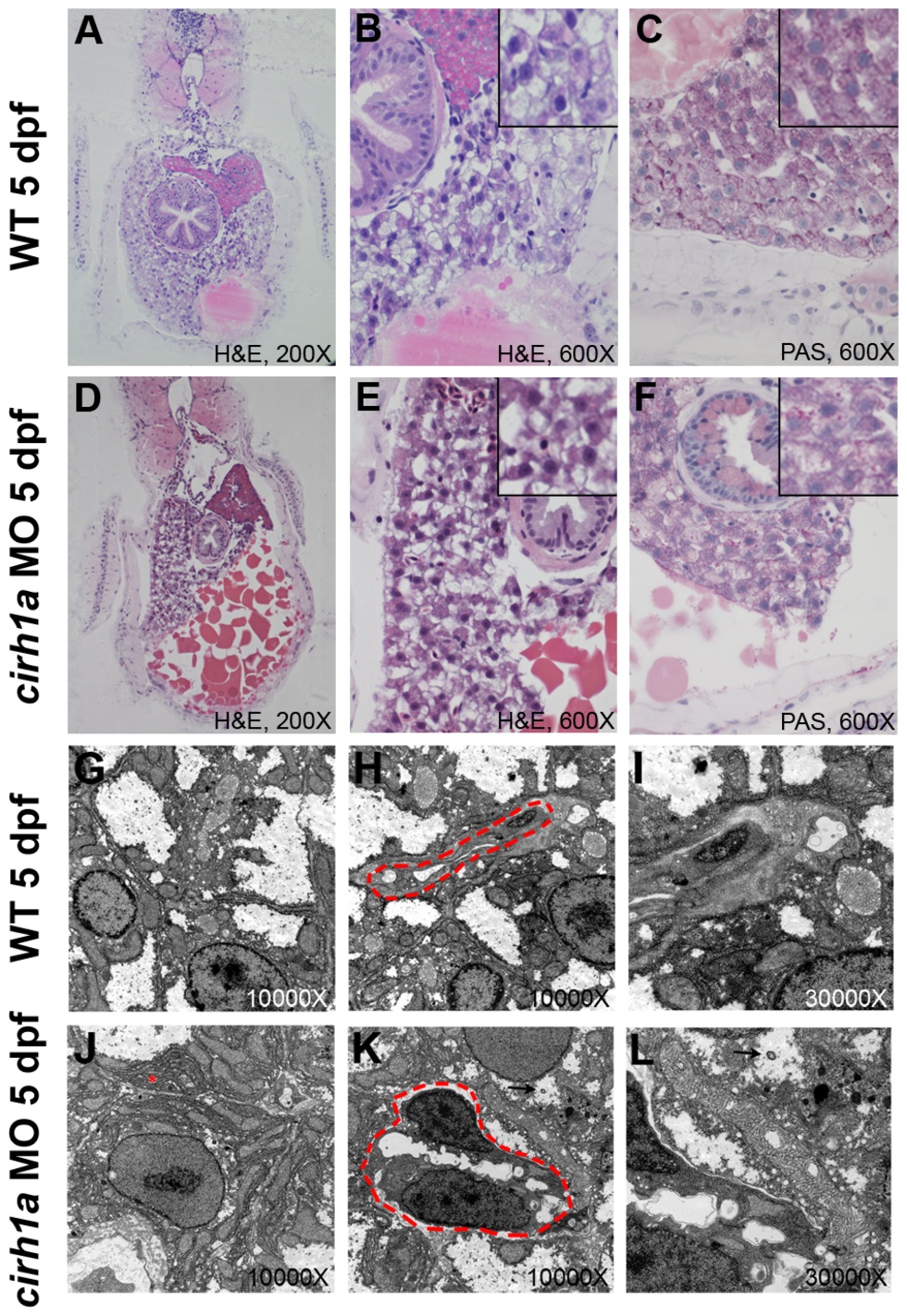
Fig. 3
Liver histology and ultrastructure in Cirhin-deficient larvae.
(A-F) Liver histology of wild-type (A-C) and Cirhin-deficient (D-F) larvae is indistinguishable, although liver size and yolk consumption are decreased in Cirhin-deficient larvae as a result of mild developmental delay (D-E). Insets in B-C, E-F are high-magnification views of associated panel. (G-L) Transmission electron microscopy of wild-type (G-I) and Cirhin-deficient (J-L) larvae. Compared to wild-type, Cirhin-deficient hepatocytes have increased rough endoplasmic reticulum (J, red asterisk) and occasional cytoplasmic lamellations consistent with bile (K-L, black arrowheads). Biliary cells are outlined by red dashed lines and appear normal (H,K).