
|
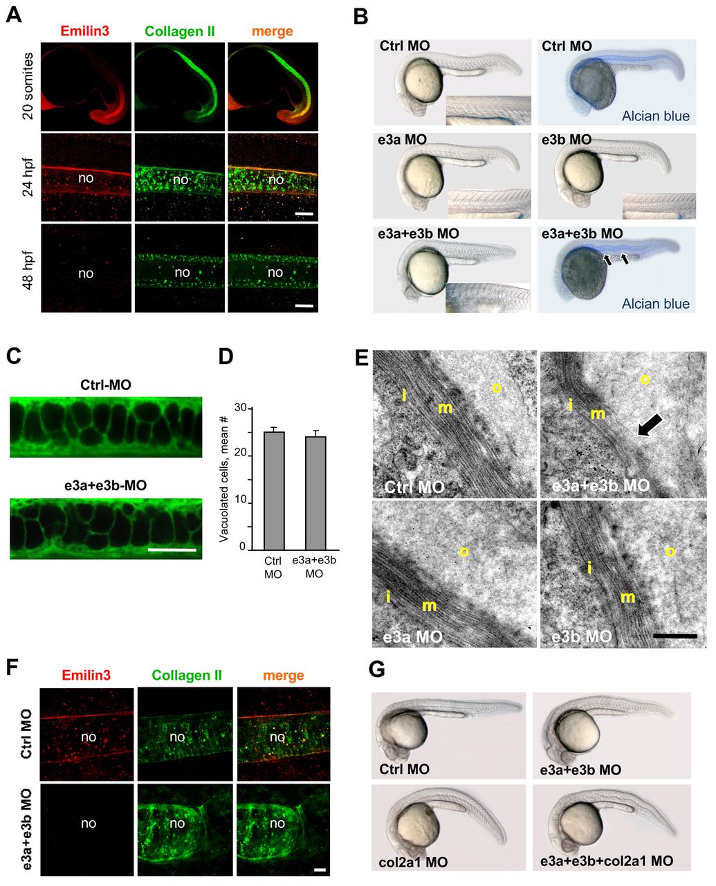
Fig. 1 Emilin3 is an essential component of the notochord sheath. (A) Lateral views of zebrafish embryos after immunofluorescence for Emilin3 and collagen II. Scale bars: 30 μm. (B) Lateral views of 24 hpf injected embryos. (C) BODIPY TR Ceramide staining of notochord cells in 24 hpf injected embryos. Scale bar: 50 μm. (D) Quantification of the mean number of vacuolated cells within the trunk region of control and treated notochord (not significantly different, n=10). Data are mean+s.e.m. (E) Transmission electron micrographs of the notochord sheath in transverse trunk sections of 30 hpf injected embryos. The arrow points at the medial layer of the notochord basement membrane, which is thinner and partially disorganized in Emilin3 double morphants. Scale bar: 500 nm. (F) Immunofluorescence for Emilin3 and collagen II in 24 hpf injected embryos. Scale bar: 10 μm. (G) Lateral views of 24 hpf injected embryos. col2a1 MO, col2a1 morpholino; Ctrl MO, control morpholino; e3a MO, emilin3a morpholino; e3b MO, emilin3b morpholino; i, inner layer; m, medial layer; no, notochord; o, outer layer.