
|
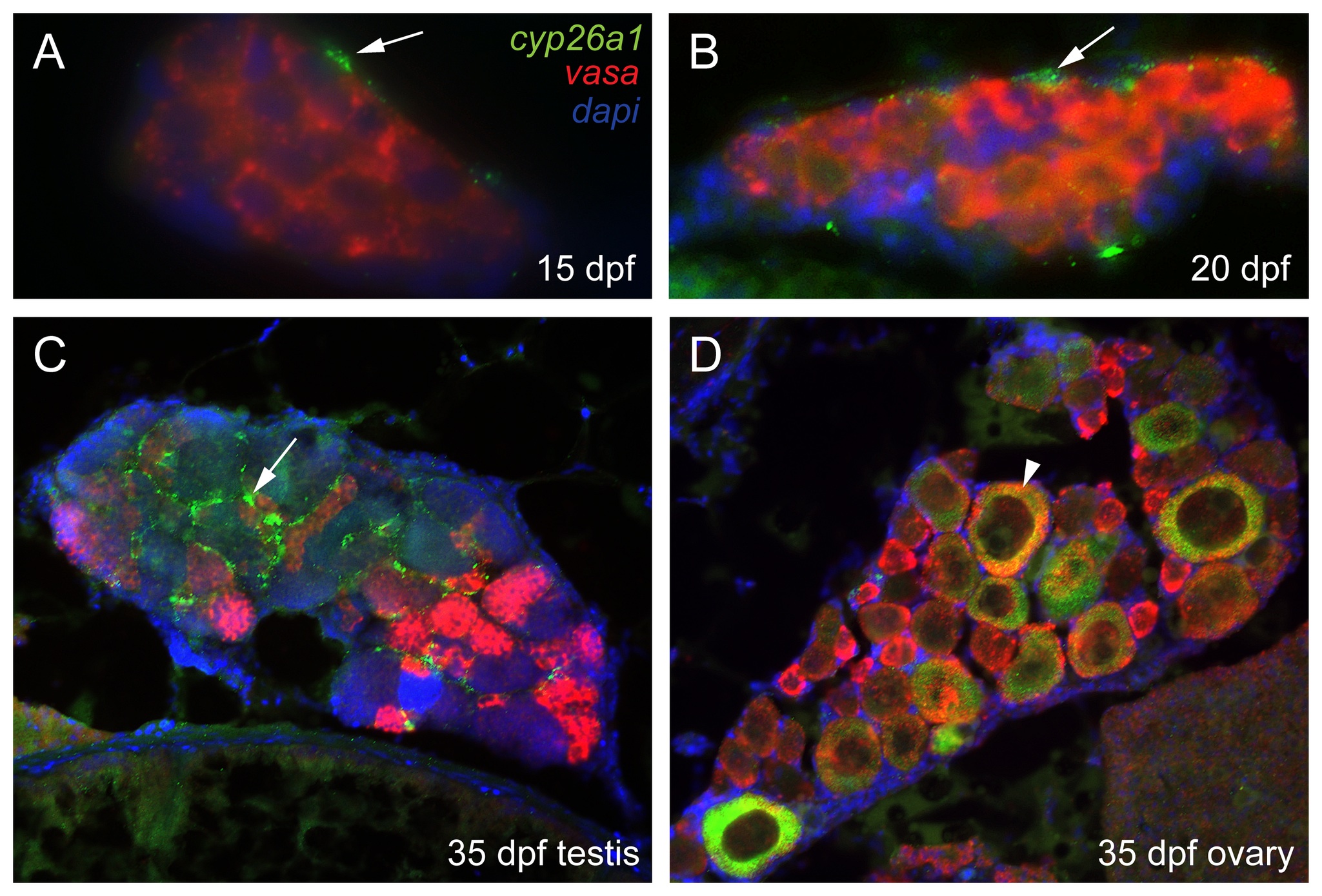
Fig. 5 Three color fluorescent detection of cyp26a1 and vasa expression during zebrafish gonad development.
Fluorescence detection of cyp26a1 (green) expression and vasa (red) expression by double in situ hybridization on gonad sections at the bipotential gonad stage at 15 dpf (A: n=1) and 20 dpf (B: n=2), and on immature gonads developing into testes (C: n=1) or ovaries (D: n=1) at 33 dpf. Cyp26a1 expression occurs in somatic cells and does not co-localize with the germ cell marker vasa in bipotential gonads (A,B). In immature testes, cyp26a1 expression was also expressed in somatic cells (arrow) and not in germ cells (C). In immature ovaries, however, cyp26a1 was expressed in large oocytes that had reached diplotene (arrowhead in D).