
Image
|
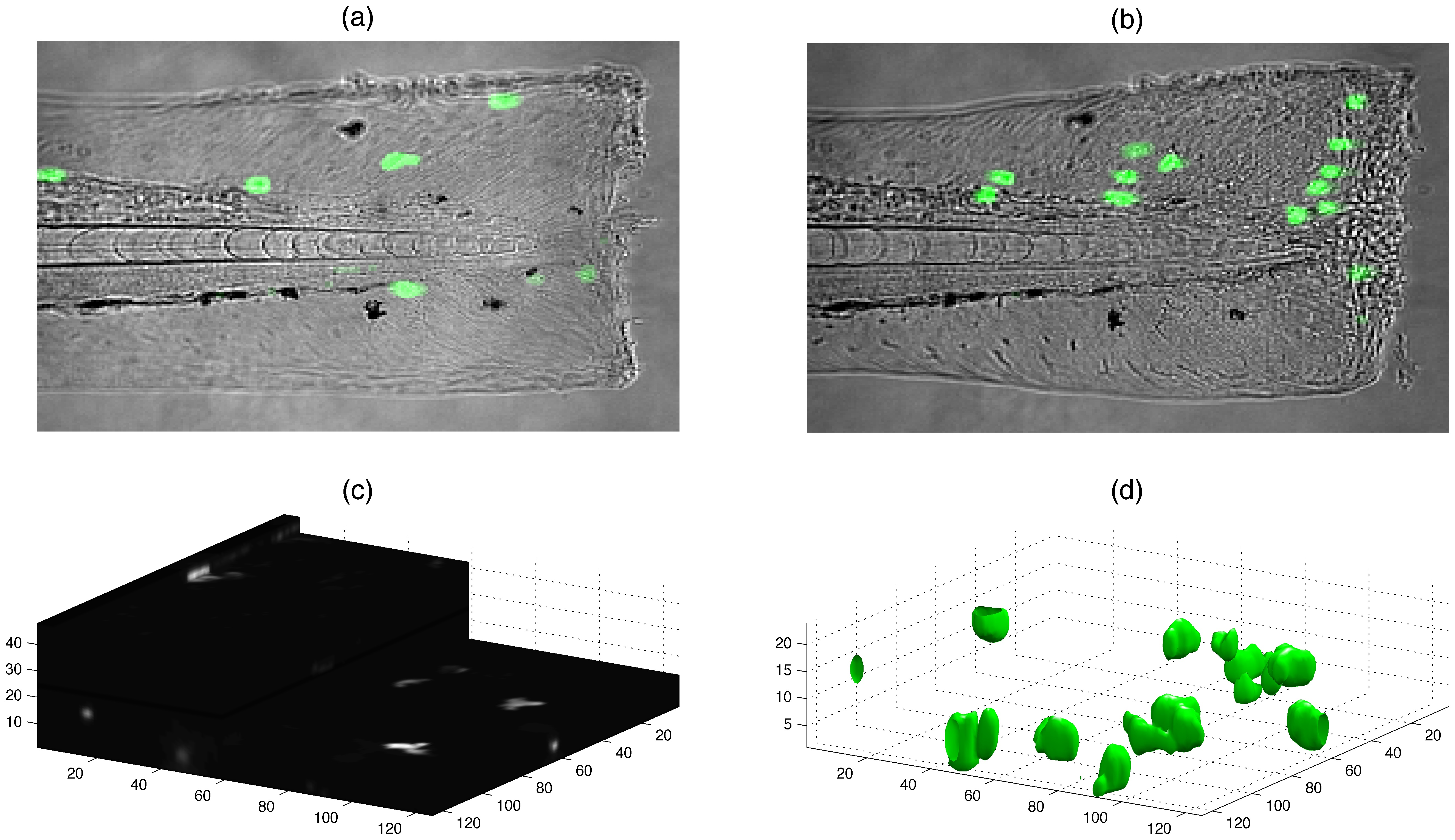
Figure Caption
Fig. 1 Visualisation of neutrophils in zebrafish.
(a,b) Fluorescent neutrophils (bright uniform regions) migrate towards the site of injury (right) in the tail-fin of a zebrafish larva (differential interference contrast (DIC)) at 3 dpf. (c) One time point of 48 slices each of 1024×1024 pixels. (d) Neutrophils rendered as 3D surfaces.
Figure Data
Acknowledgments
This image is the copyrighted work of the attributed author or publisher, and
ZFIN has permission only to display this image to its users.
Additional permissions should be obtained from the applicable author or publisher of the image.
Full text @ PLoS One