
|
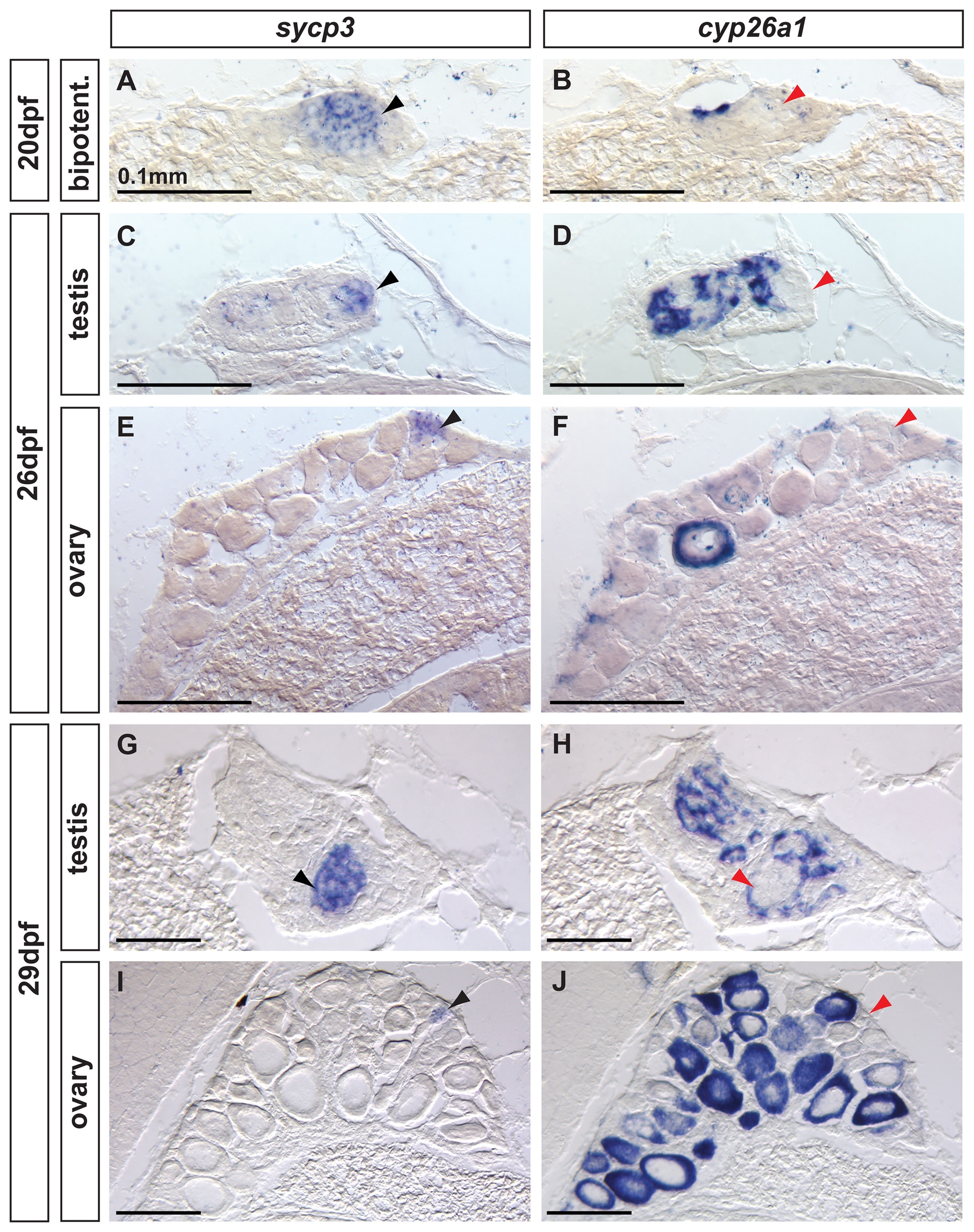
Fig. 8 Complementary expression of the meiotic recombination marker sycp3 and cyp26a1 in developing gonads.
In bipotential gonads at 20 dpf (A,B: n=8), germ cells expressed the meiotic recombination marker sycp3 (black arrowhead in A) in a non-overlapping pattern with cyp26a1 expression, which was mostly restricted to the dorsal part of the gonad (revealing that sycp3-expressing cells did not express cyp26a1 (red arrowhead in B). Expression of the meiotic marker sycp3 was detected in bipotential gonads of all animals analyzed (A, n=8) suggesting that some germ cells entered meiosis in all juveniles regardless of their definitive sex. In differentiating testes at 26 dpf (C,D: n=2) and 29 dpf (G, H: n=2), islands of germ cells that expressed sycp3 (black arrowheads in C, G) were found in an area in which RA was likely not degraded due to lack of cyp26a1 expression (red arrowheads in D,H). In contrast, in differentiating ovaries at 26 dpf (E, F: n=2) and 29 dpf (I, J: n=2), sycp3 was expressed in small germ cells (black arrowheads in E, I) that did not express cyp26a1 (red arrowheads in F, J). The expression of cyp26a1 was restricted to the ooplasm of oocytes that reached diplotene stage and entered in meiotic arrest (F,J). Scale bar: 0.1mm.