
Image
|
Figure Caption
Fig. S2
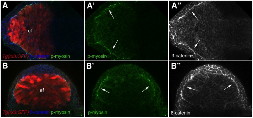
Phosphorylated light-chain-myosinII and β-catenin co-localise at the edge of the eye field.
Dorsal (A-A′′) and frontal (B-B′′) views of neural plates immunostained as shown. Arrows point at the edge of the eye field. ef: eye field.
Acknowledgments
This image is the copyrighted work of the attributed author or publisher, and
ZFIN has permission only to display this image to its users.
Additional permissions should be obtained from the applicable author or publisher of the image.
Full text @ Development