
|
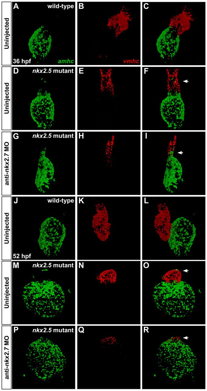
Fig. 7 Dynamic expression patterns highlight gradual transitions in cardiomyocyte identity. Two-color fluorescence in situ hybridization facilitates sensitive detection of amhc (green) and vmhc (red) expression. Confocal projections of fixed, dissected wild-type (A-C,J-L), nkx2.5 mutant (D-F,M-O) and Nkx-deficient hearts (G-I,P-R); ventral views, anterior to the top. (A-I) At 36 hpf, ectopic amhc-expressing cells are found in the ventricle in the nkx2.5 mutant heart (D-F; arrowhead) and, more extensively, in the Nkx-deficient heart (G-I; arrowhead). (J-R) At 52 hpf, ectopic amhc-expressing cells are visualized in the dramatically diminished nkx2.5 mutant ventricle (M-O; arrowhead) whereas only a tuft of residual vmhc expression remains at the pole of the Nkx-deficient heart (P-R; arrowhead).