
|
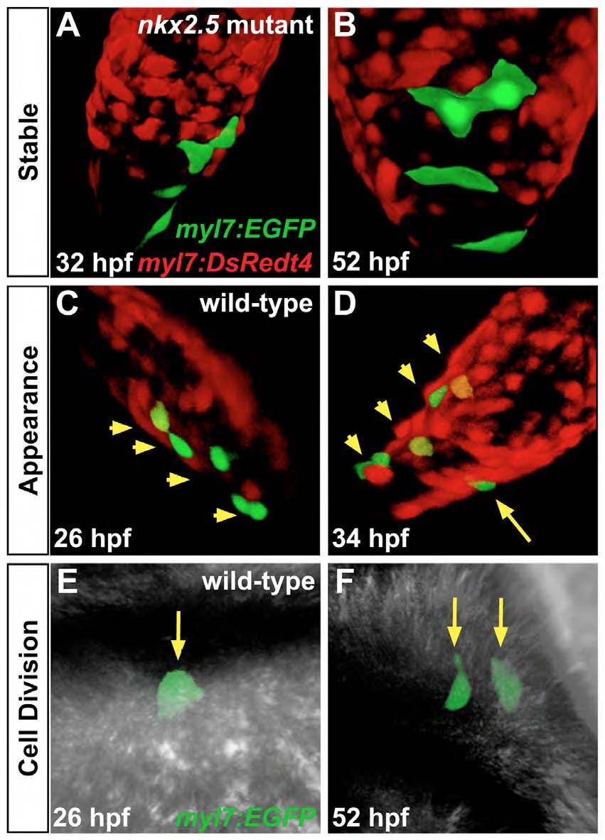
Fig. S6 Mosaic labeling tracks cardiomyocyte behavior. Confocal projections of mosaic hearts in live zebrafish embryos. Mosaic labeling was performed by injecting Tg(myl7:EGFP) plasmid into nkx2.5 mutant embryos and their wild-type siblings, both of which carried Tg(myl7:DsRedt4). Embryos with 2-6 GFPpositive cells were followed from 26 to 52 hpf, allowing for detailed analysis of cell behaviors, including division, appearance, and disappearance of cardiomyocytes. (A-F) Representative examples of observed cell behavior scenarios. The observed frequency of occurrence of each scenario in wild-type and nkx2.5 mutant embryos is provided in Table S1. (A,B) Most cells remained stable during the tracking period; as shown in this example, 4 GFP-positive cells in the atrium of a nkx2.5 mutant heart exhibit stable morphology and orientation. (C,D) In some embryos, new GFP-positive cells appeared during the tracking period; in this example, a new GFP-positive cell (yellow arrow in D) appears in a wild-type atrium. Given the distance between the new GFPpositive cardiomyocyte and the 4 originally labeled cells (yellow arrowheads in C and D), the new cell is not likely to be the product of cell division. Instead, newly appearing cells most likely reflect delayed initiation of myl7 expression in late-differentiating, SHFderived cardiomyocytes (de Pater et al., 2009; Hami et al., 2011; Lazic and Scott, 2011; Zhou et al., 2011). (E,F) Cell division was occasionally observed; in this example, a single cardiomyocyte (yellow arrow in E) in a wild-type embryo gives rise to two daughter cells (yellow arrows in F).