
Fig. 6
- ID
- ZDB-IMAGE-131218-23
- Genes
- Publication
- Sittaramane et al., 2013 - The PCP protein Vangl2 regulates migration of hindbrain motor neurons by acting in floor plate cells, and independently of cilia function
- All Figures
- Figures for Sittaramane et al., 2013
|
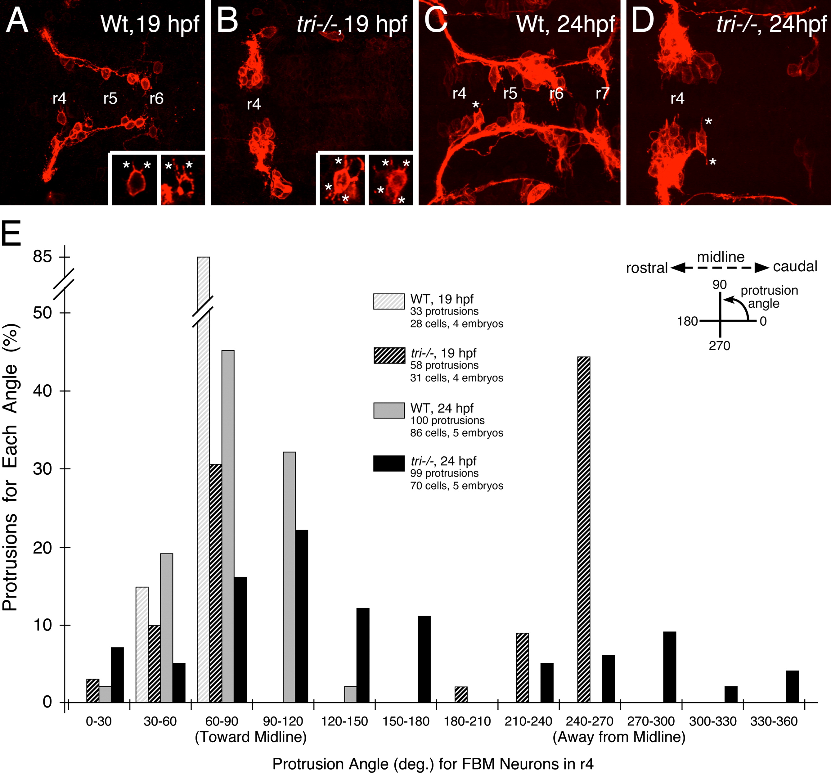
Fig. 6 Loss of orientated FBM neuron protrusions in vangl2 mutants. (AD) Dorsal views of Tg(zCREST:mRFP) hindbrains processed for RFP immunostaining. In wildtype embryos (A,C and insets), protrusions (asterisks) generated by FBM neurons in r4 are oriented toward the midline. In vangl2 mutant (tri/) embryos (B,D and insets), the protrusions of FBM neurons in r4 are oriented in various directions, with a large number directed away from the midline. (E) Quantification of the orientation of protrusions of FBM neurons in r4 of wildtype and vangl2 mutant embryos. Whereas the orientation angles for all wildtype protrusions at 19 and 24 hpf are limited to the quadrants directed toward the midline, a large number of protrusions of mutant neurons are directed 180 degrees away from the midline, especially at 19 hpf (significant difference from wildtype at p<0.001; Watson–Williams F-test).
Reprinted from Developmental Biology, 382(2), Sittaramane, V., Pan, X., Glasco, D.M., Huang, P., Gurung, S., Bock, A., Li, S., Wang, H., Kawakami, K., Matise, M.P., and Chandrasekhar, A., The PCP protein Vangl2 regulates migration of hindbrain motor neurons by acting in floor plate cells, and independently of cilia function, 400-412, Copyright (2013) with permission from Elsevier. Full text @ Dev. Biol.