
|
Fig. S10
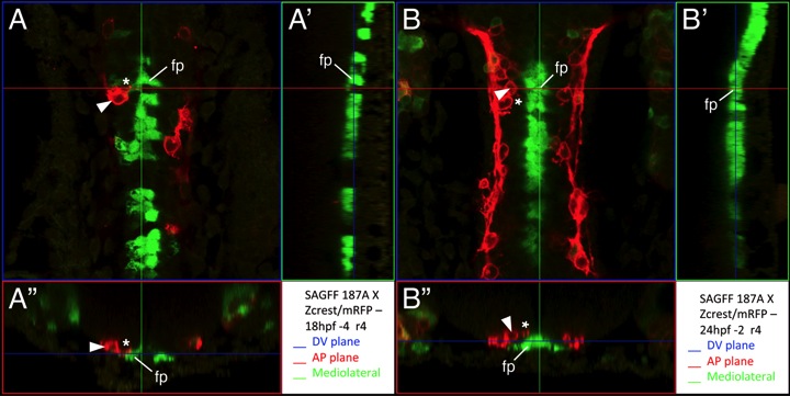
Some FBM neuron protrusions contact floor plate cells
Confocal projection views of the hindbrain of SAGFF187A aka Tg(Fp:Gal4FF); Tg(UAS:GFP); Tg(zCREST:mRFP) embryos stained with anti-RFP antibody to label FBM neurons (red) and anti- GFP antibody to label UAS:GFP-expressing cells (green). (A, B) Dorsal views at the levels indicated by blue lines in A′′ and B′′. Protrusions (asterisks) from FBM neurons (arrowheads) are very close to or in contact with floor plate cells (fp). (A′,B′) Sagittal views (mediolateral projections) at the levels indicated by green lines in A and B. (A′′,B′′) Cross-sectional views at the anterior-posterior levels indicated by red lines in A and B. The protrusions from FBM neurons are touching or in apposition to the floor plate cells. We restricted our analysis to FBM neurons located in r4/r5, and found that 34/52 protrusions (44 neurons in 8 embryos) contacted floor plate cells.
Reprinted from Developmental Biology, 382(2), Sittaramane, V., Pan, X., Glasco, D.M., Huang, P., Gurung, S., Bock, A., Li, S., Wang, H., Kawakami, K., Matise, M.P., and Chandrasekhar, A., The PCP protein Vangl2 regulates migration of hindbrain motor neurons by acting in floor plate cells, and independently of cilia function, 400-412, Copyright (2013) with permission from Elsevier. Full text @ Dev. Biol.