
|
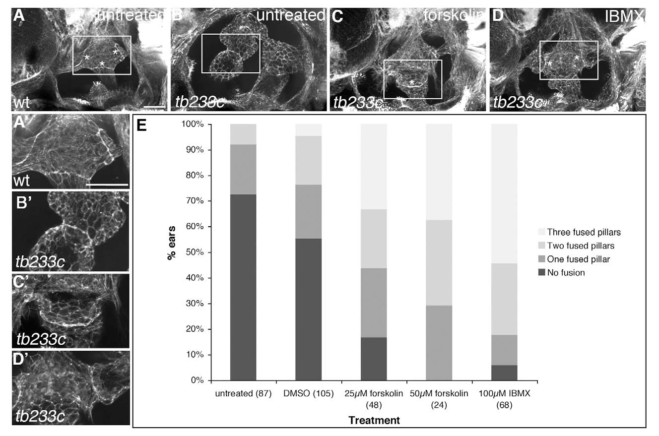
Fig. S5 Treatment with cAMP agonists can rescue fusion plate formation in lauscher mutant embryos. (A-D′) Confocal images of ears stained with FITC-phalloidin (marking F actin) to show morphology of the canal projections and pillars. Concentrations of actin mark the fusion plates (shown at higher magnification in A′-D′). (A, A′) Wild-type ear. Three fusion plates are clearly visible (asterisks). (B,B′) Untreated ear from a homozygous tb233c mutant. Canal tissue is very disorganised. Although projections are touching in this ear, analysis of the z-stack indicated that this was not a true fusion plate. (C-D′) Treatment of homozygous tb233c embryos with either 50μM forskolin or 100μM IBMX can restore pillar formation. Fusion plates appear relatively normal. (E) Quantitation of the number of fusion plates present. Data were analysed with a 433 chi-square contingency table (DMSO, forskolin) or a 432 chi-square contingency table (DMSO, IBMX); P<0.001 for both drugs. N numbers are shown in parentheses. Lateral views. Scale bars: A-D 50μm; A′-D′μ.