
Image
|
Figure Caption
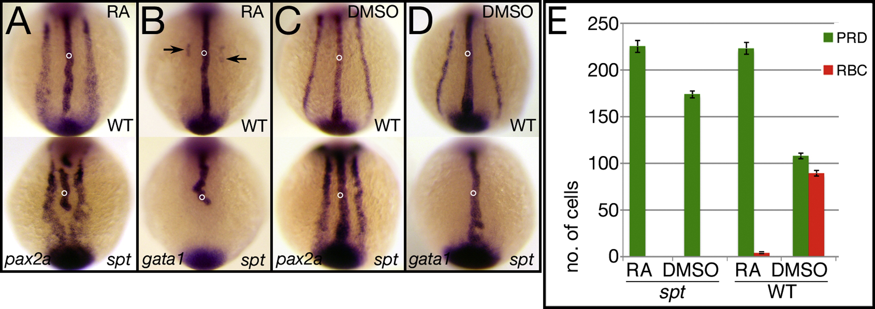
Fig. S3 Transient exposure to RA gives results similar to overexpression of Fgf8a. (A–E) Exposure at 6 h to 1 μM RA for 1 h. (A–D) in situ analysis at 12 h. Control siblings were exposed to just the carrier (5% DMSO). pax2a expression identifies pronephric precursors (PRD), and gata1 expression identifies red blood precursors (BLD). Embryos were also probed with ntl (open circle). (E) Quantification of precursor cells at 12 h. Graphs show the average number of cells per fate and the standard error. Embryos are shown from a posterior view.
Acknowledgments
This image is the copyrighted work of the attributed author or publisher, and
ZFIN has permission only to display this image to its users.
Additional permissions should be obtained from the applicable author or publisher of the image.
Reprinted from Developmental Biology, 383(1), Warga, R.M., Mueller, R.L., Ho, R.K., and Kane, D.A., Zebrafish Tbx16 regulates intermediate mesoderm cell fate by attenuating Fgf activity, 75-89, Copyright (2013) with permission from Elsevier. Full text @ Dev. Biol.