
|
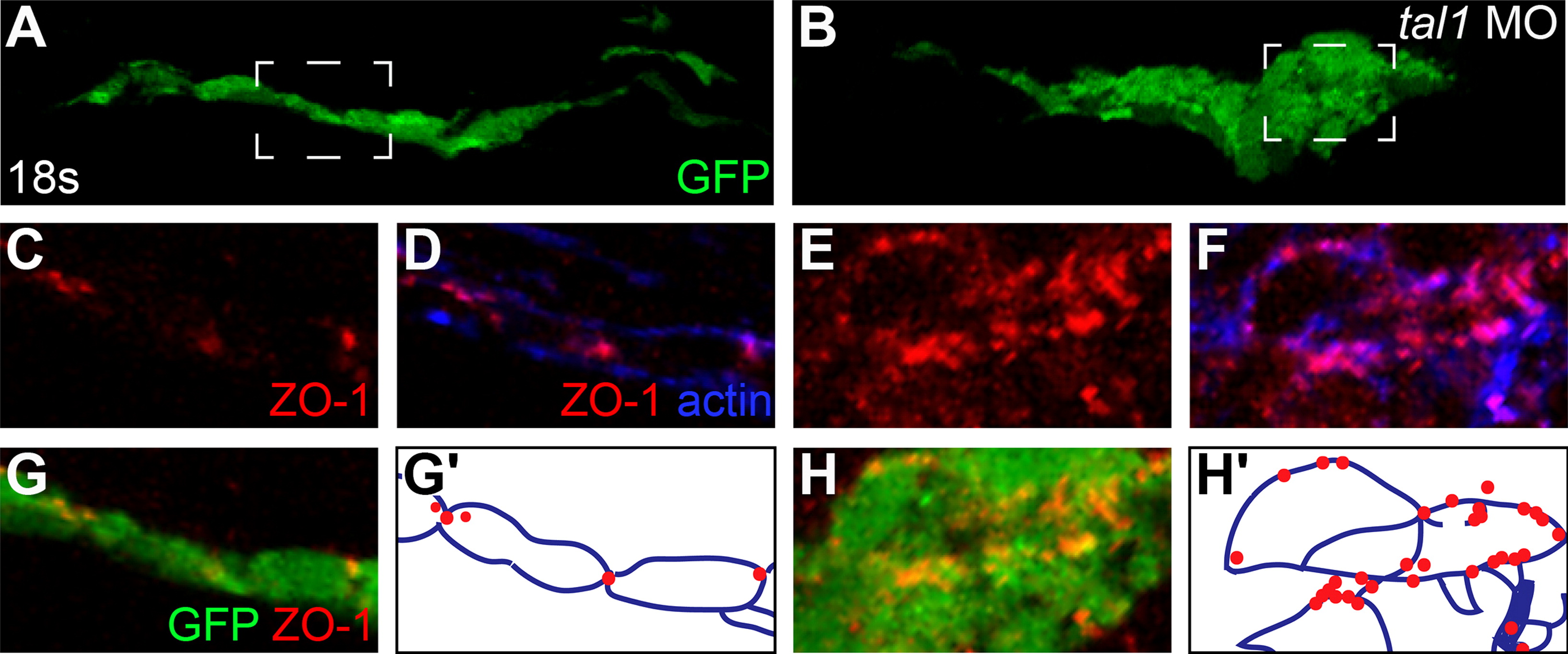
Fig. 6 Endocardial intercellular junctions are disorganized during early endocardial morphogenesis in tal1-deficient embryos. (A–H) Transverse sections of wild-type (A, C, D, and G) and tal1-deficient embryos (B, E, F, and H), dorsal to the top, at 18s. Immunofluorescence detects localization of ZO-1 (red) (C–H) and F-actin (blue) (D and F); in addition, an anti-GFP antibody allows visualization of Tg(kdrl:GRCFP) expression (green) in the endocardium (A, B, G, and H). The boxed areas in (A and B) are highlighted in (C–H). (A) Wild-type endocardium forms a single-layered sheet with ZO-1 localized to discrete puncta at cell-cell contacts (C, D, and G). (B) tal1-deficient endocardium is multi-layered and disorganized, and ZO-1 is mislocalized in multiple puncta along the endocardial cell boundaries (E, F, and H). (G′ and H′) Cartoons indicate regions of ZO-1 (red) and F-actin (blue) localization within the endocardium.
Reprinted from Developmental Biology, 383(2), Schumacher, J.A., Bloomekatz, J., Garavito-Aguilar, Z.V., and Yelon, D., tal1 regulates the formation of intercellular junctions and the maintenance of identity in the endocardium, 214-226, Copyright (2013) with permission from Elsevier. Full text @ Dev. Biol.