
|
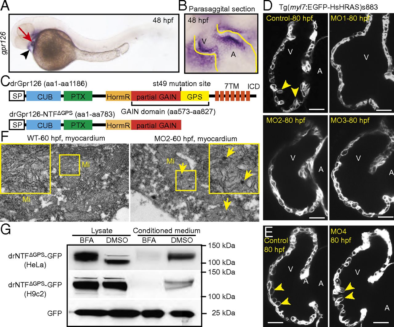
Fig. 3 Gpr126 knockdown leads to cardiac abnormalities in zebrafish. (A) Lateral view of a 48-hpf zebrafish embryo after whole-mount in situ hybridization showing gpr126 expression in heart (red arrow) and pericardium (black arrowhead). (B) Parasagittal section confirming cardiac expression of gpr126. (C) Schematic representation of zebrafish full-length Gpr126 (drGpr126) and its NTF part up to the st49 mutation site (drGpr126-NTFΔGPS). (D and E) Confocal sections of hearts from control- and morpholino-injected Tg(myl7:EGFP-HsHRAS)s883 embryos at 80 hpf. In full-length Gpr126-depleted animals (MO1-3) but not in Gpr126-CTF–depleted (MO4) morphants, trabeculation (yellow arrowheads) is perturbed. (Scale bar: 20 μm.) (F) Transmission electron micrographs at 60 hpf reveal that morphants contain elongated Mi with more branched cristae and electron dense precipitates (arrows) compared with WT siblings. (G) Western blot analysis of lysates and conditioned medium from cells overexpressing C-terminal GFP-tagged drGpr126-NTFΔGPS or GFP. Note that both GFP-tagged NTFΔGPS (predicted band size: 113 kDa) and GFP (predicted band size: 27 kDa) were detected in conditioned medium. However, secretion of GFP-tagged NTFΔGPS but not GFP was inhibited by BFA (a blocker of classical trans-Golgi secretory pathway). A, atrium; V, ventricle.