
|
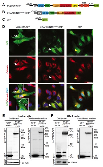
Fig. S6 The NTF part of the st49 mutant form of zebrafish Gpr126 is an independent, stable, secreted protein. (A and B) Schematic depiction of the C-terminal GFP-tagged drGpr126 (A) and drGpr126-NTFΔGPS (B). ICD, intracellular domain. (C) GFP vector used as control. (D) Immunofluorescence analysis of HeLa cells stained for GFP (green) and calnexin (red) after transfection with constructs described in A–C. Nuclei were stained with DAPI (blue). drGpr126-GFP localized to the plasma membrane (white arrowheads) and colocalized with calnexin (arrows), an endoplasmic reticulum marker protein. drGpr126-NTFΔGPS-GFP was not detected at the plasma membrane but colocalized with calnexin (arrows). GFP was predominantly expressed in the nucleus (yellow arrowheads). (E and F) Western blot analysis of cell lysates and conditioned medium from HeLa (E) and H9c2 cells (F) after transfection with C-terminal GFP-tagged drGpr126-NTFΔGPS or GFP. Note that GFP-tagged NTFΔGPS (predicted band size: 113 kDa) and GFP (predicted band size: 27 kDa) were detected in both lysate and conditioned medium. Gapdh was used as loading control for cell lysates.