
|
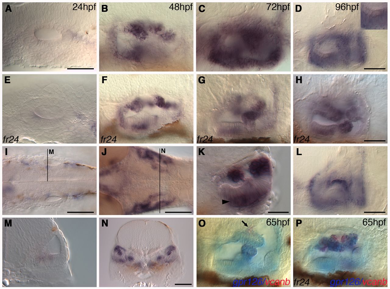
Fig. 6 Expression of gpr126 in wild-type and lauscher mutant ears. (A-H) Expression of gpr126 mRNA in the ear at 24-96 hpf in wild-type (A-D) and fr24 mutant embryos (E-H). Strongest expression is in canal projections prior to fusion (48-72 hpf), with some expression remaining at 96 hpf. Inset in D: higher magnification showing expression in the anterior macula supporting cell layer. (I,M) Wild-type expression of gpr126 at 26 hpf in the anterior macula (I, dorsal view; M, transverse section). (J,N) Expression at 48 hpf shows stronger staining in the projections and sensory patches (J, dorsal view; N, transverse section). (K) Expression in sensory patches at 72 hpf is restricted to supporting cells (arrowhead) (transverse section; see also inset in D). There is strong expression in the projections. (L) Alternative focus view of D, showing residual expression in the lateral projection at 96 hpf. (O,P) Expression of gpr126 (blue) and vcanb (red) in the projections of wild-type and fr24 mutant embryos at 65 hpf. In wild-type embryos (O), there is co-expression (purple) in the recently fused ventral pillar; vcanb is downregulated in the lateral projection and in the anterior and posterior pillars (out of focus), whereas gpr126 is expressed at reduced levels. In fr24 mutants (P), vcanb and gpr126 are co-expressed in the unfused projections. Expression of vcanb persists in the dorsolateral septum, which does not express gpr126, in both wild-type and mutant embryos (O, arrow). Scale bars: in A, 50 μm for A-C,E-G,M,O,P; 50 μm in D,H,L; 25 μm in K; 100 μm in I,J,N.