
|
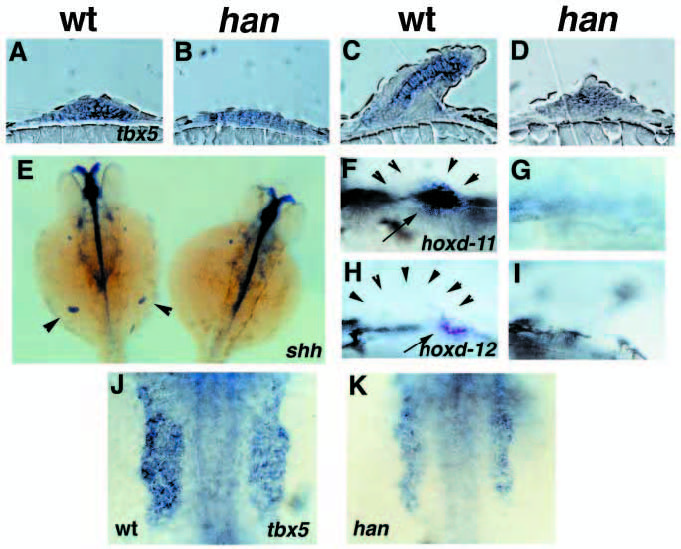
Fig. 3 Pectoral fin defects in hans6 mutants. (A-D) Longitudinal sections through pectoral fin buds after in situ hybridization for tbx5 expression, anterior to the left. (A) At 32 hpf, a fin bud is forming in wild-type embryos. (B) hans6 mutants exhibit a delay in fin bud formation as well as a reduction in tbx5 expression at this stage. (C) In 48 hpf wild-type embryos, the pectoral fin is elongating and a chondrogenic condensation is forming. tbx5 expression is highest in the chondrogenic portion of the pectoral fin at this stage. (D) In hans6 mutants, a small undifferentiated fin bud expresses a reduced level of tbx5. (E) Dorsal views, anterior to the top, of a wild-type embryo (left) and a hans6 mutant (right) at 36 hpf. Embryos are golden homozygotes. shh expression is visible in the ZPA of each pectoral fin bud in wild-type embryos (arrowheads) but not in hans6 mutants. (F-I) Lateral views, anterior to the left, of pectoral fin buds from wild-type (F,H) and hans6 mutant (G,I) siblings at 32 hpf. (F) Wild-type embryos express hoxd-11 in a posterior portion (arrow) of the fin bud (outline indicated by arrowheads); (G) hans6 mutants never express hoxd-11 in the fin mesenchyme. (H) Wild-type embryos express hoxd-12 in a posterior portion (arrow) of the fin bud (outline indicated by arrowheads); (I) hans6 mutants never express hoxd-12 in the fin mesenchyme. (J,K) Dorsal views, anterior to the top, of the pectoral fin-forming region of the LPM in wild-type (J) and hans6 mutant (K) embryos at the 16-somite stage (17 hpf). The domain of tbx5 expression is expanded in wild-type embryos, but not in hans6 mutants.