
|
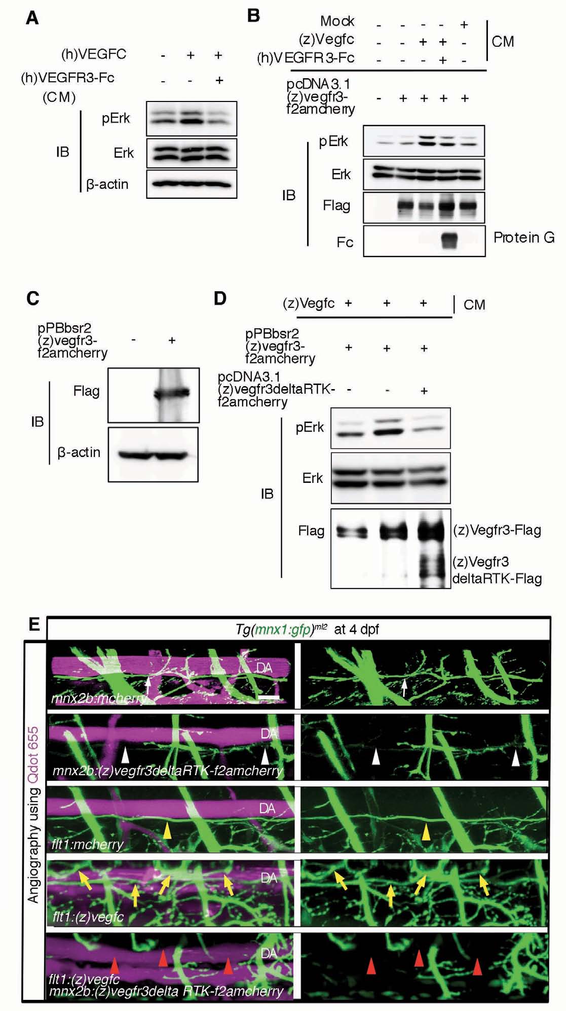
Fig. S5 Impaired parallel growth of motoneurons after inhibition of vegfr3 in motoneurons. (A) Immunoblot analyses with the antibodies indicated at the left using cell lysates of HUVECs treated with recombinant human (h)VEGFC together with or without a truncated mutant of (h)VEGFR3 tagged with human IgG Fc, (h)VEGFR3-Fc, prepared from the conditioned medium (CM) of 293T cells transfected with the plasmids expressing (h)VEGFR3-Fc. (B) Immunoblot analyses with the antibodies indicated at the left using cell lysates of 293T cells transfected with or without the plasmid expressing zebrafish (z)Vegfr3 tagged with Flag followed by 2A peptide and mCherry (pcDNA3.1(z)vegfr3-f2amcherry) and treated with the CM indicated at the top. Bottom panel, precipitates on protein G and subjected to immunoblot with anti-human immunoglobulin. (C) Immunoblot analyses with the antibodies indicated at the left using the cell lysates of the parental and HEK293 cells stably transfected with pPBbsr2(z)vegfr3-f2amcherry plasmids using piggyBac transposon system. (D) Immunoblot analyses with the antibodies indicated at the left using the lysates of the cells described in C transfected with the plasmid expressing zebrafish (z)Vegfr3 lacking the cytoplasmic domain tagged with Flag followed by 2A peptide and mCherry (pcDNA3.1(z)vegfr3deltaRTKf2amcherry) and treated with the conditioned medium (CM) as indicated at the top. (E) 3D-rendered confocal stack images (lateral view) of Tg(mnx1:gfp)ml2 embryos injected with the plasmids indicated at the bottom of each panel at 4 dpf. Blood vessels were visualized by injecting Quantum (Q) dot 655 into the blood vessels. Left panels, merged images of GFP and Qdot 655 images; right panels, GFP images. White arrows and yellow arrowheads indicate parallel growth (PG) of motoneuron axons beneath the DA. White arrowheads and red arrowheads denote the impairment of PG of motoneuron axons and DA. Yellow arrows indicate the increased branches of motoneuron axons. Scale bar: 25 μm. DA, dorsal aorta.