
|
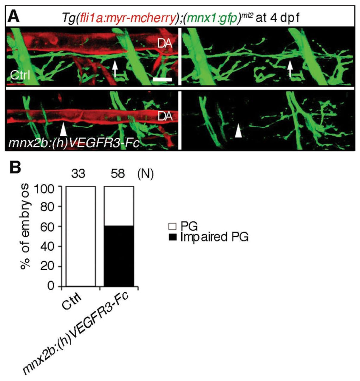
Fig. 7 Inhibition of Vegfr3 in the motoneuron results in impairment of alignment of the motoneuron axons and dorsal aorta. (A) 3D-rendered confocal stack images (lateral view) of Tg(fli1a:myr-mcherry);(mnx1:gfp)ml2 embryos (upper panel) and those transiently expressing (h)VEGFR3-Fc in the motoneurons under the control of the mnx2b promoter by Tol2-mediated gene transfer (lower panels). Left panels, merged images of mCherry and GFP; right panels, GFP images. Arrows indicate the motoneuron axon beneath the dorsal aorta (DA). Arrowheads indicate the impairment of parallel growth of motoneuron axon with the DA. Scale bar: 25 μm. (B) Quantitative analyses of the impaired parallel growth (PG) of the embryos grouped as in A. The number (n) of the embryos observed indicated at the top.