
|
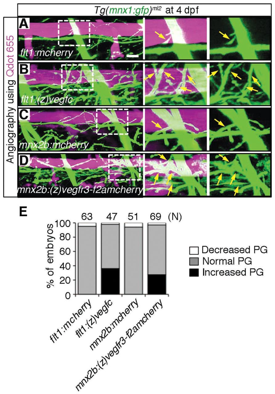
Fig. 5 Overexpression of either Vegfc in the dorsal aorta or Vegfr3 in the motoneurons enhances axon branches of motoneurons over the dorsal aorta. (A-D) 3D-rendered confocal stack images (lateral view) of Tg(mnx1:gfp)ml2 embryos transiently expressing the molecules indicated at the bottom left of the left-hand panels. The embryos were intravascularly injected with Qdot 655. Left panels, merged images of GFP and Qdot 655 images; center panels, enlarged image of boxed region of the left panels; right panels, enlarged image of boxed region of the left panels showing GFP signal only. (A) Tg(mnx1:gfp)ml2 embryo transiently expressing mCherry in arteries under the control of the flt1 promoter by Tol2-mediated gene transfer. Scale bar: 25 μm. (B) Zebrafish expressing (z)Vegfc similar to A. (C) Embryo transiently expressing mCherry in motoneurons under the control of the mnx2b promoter by Tol2-mediated gene transfer. (D) Embryo transiently expressing zebrafish (z)Vegfr3 tagged with Flag (F) followed by 2A peptide and mCherry [(z)Vegfr3-F2AmCherry] similar to C. Arrows indicate branches of motoneurons. (E) Quantitative analyses of branching of axons over the DA. The number of the embryos showing the increased, normal or decreased parallel growth (PG) divided by the total number (n) of embryos counted (indicated at the top) is expressed as percentage of embryos.