
|
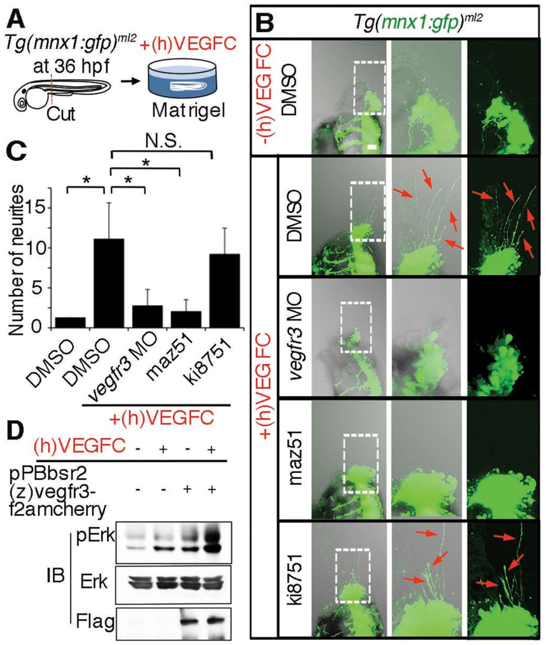
Fig. 4 Motoneurons extend neurites in response to recombinant VEGFC. (A) Schematic of an ex vivo assay for examining neurite sprouting from motoneurons. Briefly, the Tg(mnx1:gfp)ml2 embryos were cut at the dorsoventral line between the main yolk and the rostrally extended yolk tube and was embedded in matrigel with or without recombinant human vascular endothelial growth factor-C, (h)VEGFC. (B) z-stack confocal images (left panel, merged image of transmitted light image and GFP image; center, enlarged image of the boxed region in the left panel; right, GFP image of the boxed region in the left panel) of Tg(mnx1:gfp)ml2 embryos incubated in matrigel. Upper panels, embryo in matrigel without (h)VEGFC; lower four panels, embryos in matrigel with (h)VEGFC and treated as indicated at the left. Red arrows indicate GFP-positive neurites from motoneurons. Anterior is at the top and dorsal to the right. (C) Quantitative analyses (ANOVA) of neurite number of the embryos grouped as in B. *P<0.05. Plotted is mean ± s.d. of more than ten embryos for each group. (D) Immunoblot analyses with the antibodies indicated at the left using lysates of parental HEK293 and those stably transfected with pPBbsr2(z)vegfr3-f2amcherry and treated with or without (h)VEGFC. A representative result of three independent experiments is shown.