
IMAGE
Fig. S2
- ID
- ZDB-IMAGE-131211-14
- Publication
- Zhen et al., 2013 - Hemogenic endothelium specification and hematopoietic stem cell maintenance employ distinct Scl isoforms
- All Figures
- Figures for Zhen et al., 2013
Image
|
Figure Caption
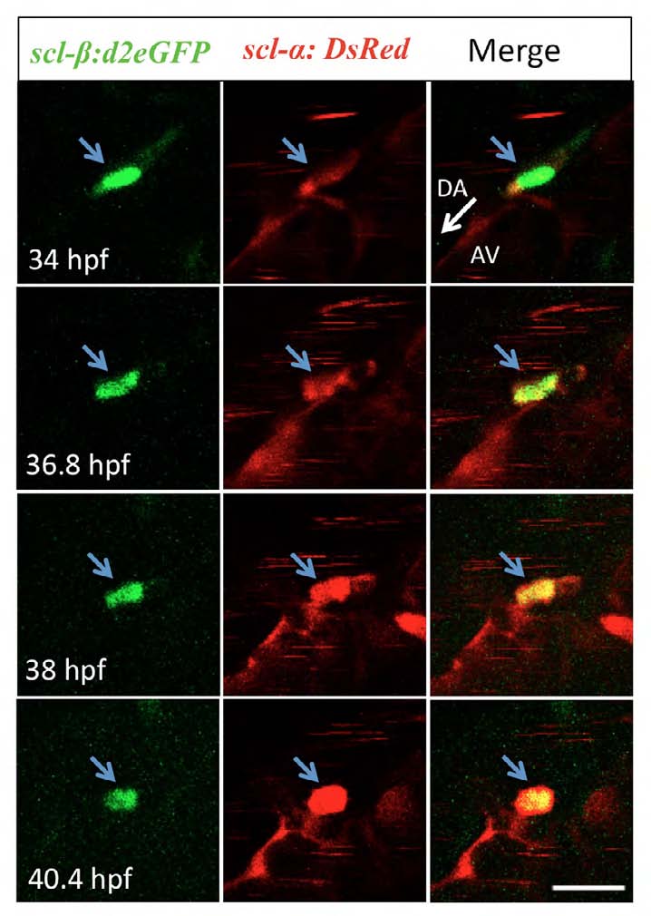
Fig. S2 scl-β:d2eGFP+ endothelial cells give rise to scl-β:d2eGFP+/scl-α:DsRed+ HSCs. Time-lapse confocal imaging of a live Tg(scl-β:d2eGFP; scl-α:DsRed) embryo between 34 and 40 hpf. Four selected time points show the stepwise transition of an scl- β:d2eGFP+ endothelial cell to an scl-β:d2eGFP+/scl-α:DsRed+ HSC via EHT (blue arrows). The intensity of DsRed signal is increased as the cell bends outwards. For each time point, d2eGFP, DsRed and merged images are presented. White arrow indicates the direction of circulation in DA. DA, dorsal aorta; AV, axial vein. Scale bar: 20 μm.
Acknowledgments
This image is the copyrighted work of the attributed author or publisher, and
ZFIN has permission only to display this image to its users.
Additional permissions should be obtained from the applicable author or publisher of the image.
Full text @ Development