
Image
|
Figure Caption
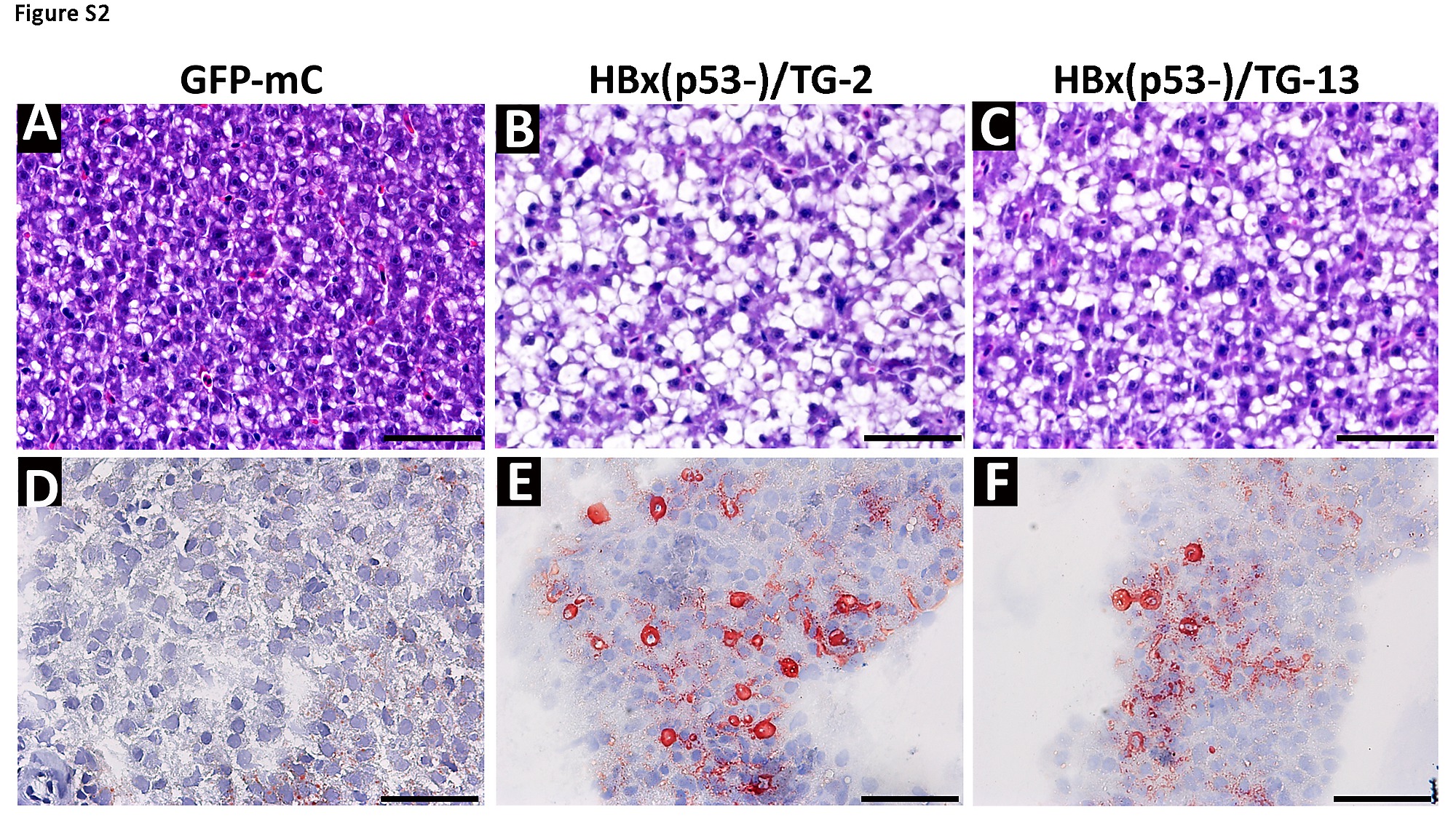
Fig. S2
Oil-red staining proved the prominent vacuoles in the cytoplasm of hepatocytes of transgenic fish exhibited lipid accumulation. (A~C) The liver samples from GFP-mC control fish and two strains of HBx transgenic fish in p53 mutant background were stained with hematoxylin-eosin after paraffin embedding. (D~E) The same liver were frozen and stained with oil red O indicated the lipid accumulated in the vacuoles in the hepatocytes. (x 400). Scale bars: 50μm.
Acknowledgments
This image is the copyrighted work of the attributed author or publisher, and
ZFIN has permission only to display this image to its users.
Additional permissions should be obtained from the applicable author or publisher of the image.
Full text @ PLoS One