
|
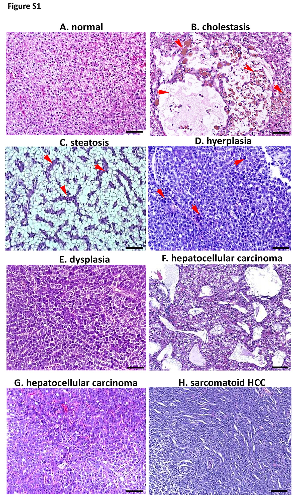
Fig. S1
Typical histological features of liver of HBx and src transgenic fish (B-H) in comparison with these of GFP-mCherry transgenic fish. (A) Liver of GFP-mCherry transgenic fish. Normal liver tissue cells arranged in neat rows, the size of the nucleus are similar, and the nuclear-cytoplasmic ratio is not too high. (B) Cholestasis: deposition yellow-green globular bilirubin pigment, arrows indicated bile and spongy vacuoles. (X200). (C) Steatosis: prominent vacuoles in the cytoplasm of hepatocytes, arrows indicated lipid droplets formed by the vacuoles. (X200). Hyperplasia and dysplasia was based on the degree of cell proliferation, also consider the whole area overall differentiation and cell differentiation, such as nuclear-cytoplasmic ratio etc. (D) Hyperplasia: disordered proliferation of atypical hepatocytes with enlarged and mildly irregular nuclei, arrows indicated single or several larger cells with higher nuclear-cytoplasmic ratio compared to surrounding adjacent normal cells (X200). (E) Dysplasia: transformed cells with enlarged nuclei and prominent nucleoli. (X200). Hepatoma cell morphology differs in texture due to the degree of differentiation, such as poorly differentiated, well differentiated, moderately differentiated etc. (F) Hepatocellular Carcinoma (HCC): hepatocellular carcinoma with marked cystic degeneration (spongiosis hepatis) (X200). (G) Hepatocellular Carcinoma (HCC): severe sheets of tumor cells with enlargement polymorphic nuclei and prominent nucleoli. (X200). (H) Sarcomatoid HCC: pleomorphic spindle tumor cells growing in haphazardly fascicular patterns. (X200).