
Image
|
Figure Caption
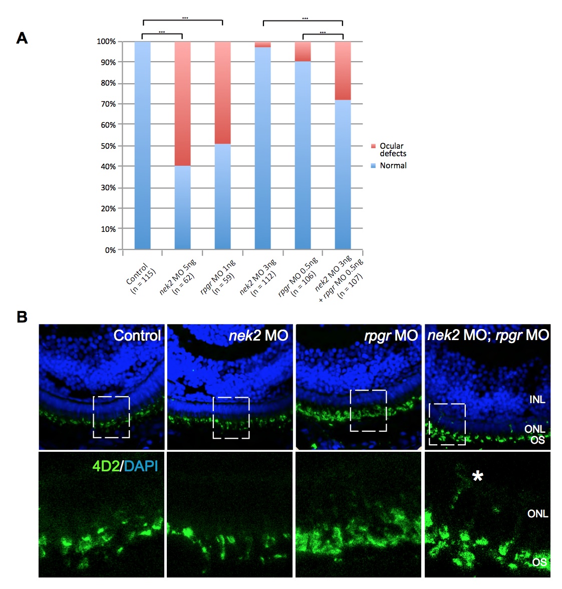
Fig. S6 In vivo functional evaluation of nek2 and rpgr deficiency in zebrafish. (A) Ocular phenotypes are quantified in control, nek2 MO, rpgr MO, and nek2 MO + rpgr MO embryos at full and sub-effective doses. (B) Immunohistochemical analyses of retinal cryosections from control, nek2 MO, rpgr MO, and nek2 + rpgr MO embryos at sub- effective doses, stained with DAPI (blue) and 4D2 antibody (green). Asterisk denotes excessive rhodopsin in the outer nuclear layer (ONL). OS, outer segment of photoreceptors; INL, inner nuclear layer.
Figure Data
Acknowledgments
This image is the copyrighted work of the attributed author or publisher, and
ZFIN has permission only to display this image to its users.
Additional permissions should be obtained from the applicable author or publisher of the image.
Full text @ Proc. Natl. Acad. Sci. USA