
Image
|
Figure Caption
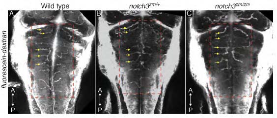
Fig. S1
Microangiography of the vasculature in notch3 mutants. Images of Fluorescein-dextran labeled vasculature of 60-63 hpf A) wild type (n=2), B) notch3zm/+ (n=10) and C) notch3zm/zm (n=6) larvae reveals comparable patterning of the hindbrain vessels at the anatomical level (compare red dashed boxed areas). Yellow arrows indicate the Central arteries (CtAs). Images are max projections of dorsal views.
Acknowledgments
This image is the copyrighted work of the attributed author or publisher, and
ZFIN has permission only to display this image to its users.
Additional permissions should be obtained from the applicable author or publisher of the image.
Full text @ Dis. Model. Mech.