
|
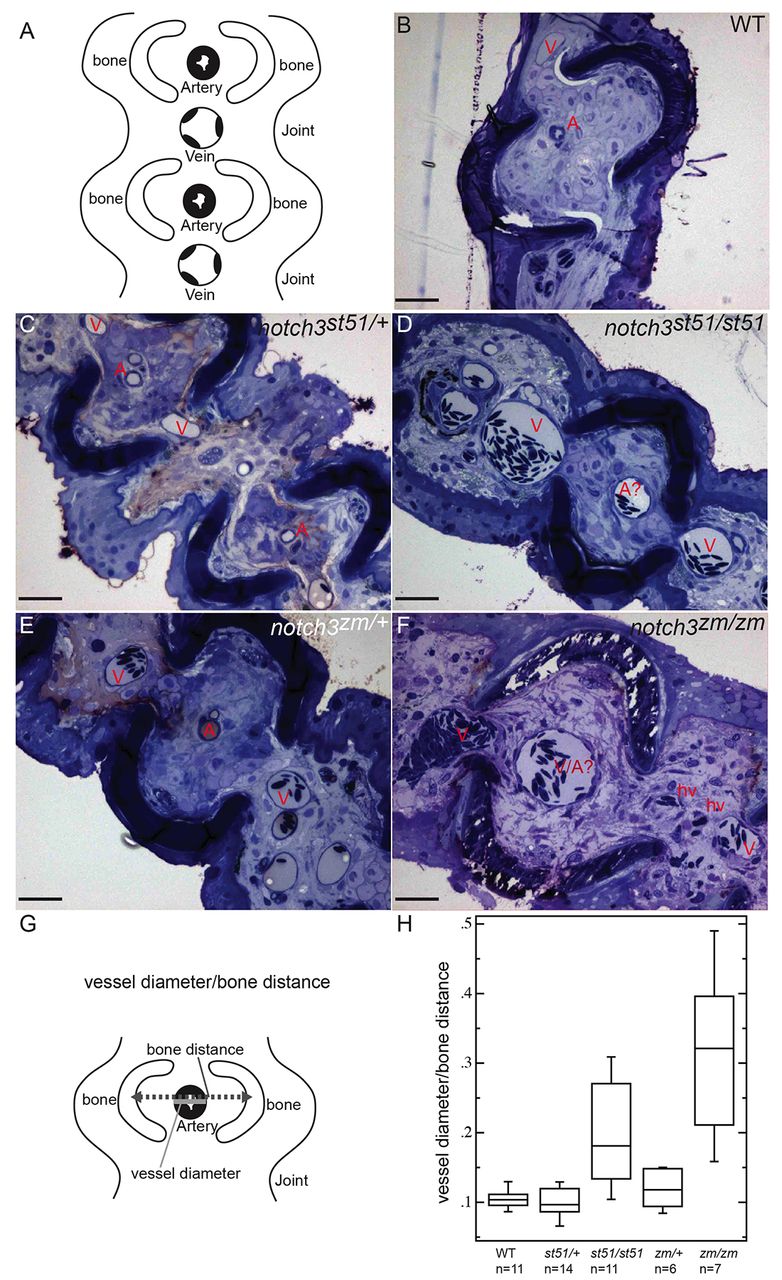
Fig. 6
Distension of vessels in notch3 mutants. (A) Schematic depicting cross-section and the ordered arrangement of arteries within the bony regions and larger caliber veins within the joints of the fin rays observed in (B) wild type, (C) notch3st51/+ and (E) notch3zm/+ heterozygotes. This pattern is moderately disrupted in (D) notch3st51/st51 mutants and severely disrupted in (F) notch3zm/zm mutants. (B–F) Toluidine-blue-stained sections. Scale bars: 50 μm. A, artery; A?, probable artery based on position; V, vein; hv, hemorrhaged vessel. (G) Schematic depicting the strategy for quantification of vessel defects (detailed in the Materials and Methods). (H) Box plot of results reveals significantly larger vessels within the bony segments of the fin of notch3 mutants compared with wild-type adults. ANOVA results: mean squares between=6.7821×10²; mean squares error=3.4565×10³; P<0.0001.