
|
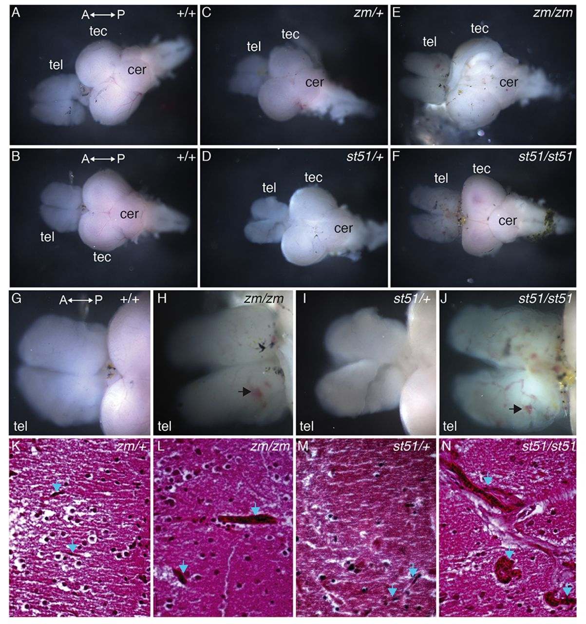
Fig. 5
Vessels are more prominent in the telencephalon of notch3 mutants compared with wild type. (A–F) Overview images of dissected adult brains. (G–J) High-magnification images of the telencephalon. Compared with (A,B,G) wild-type and (C,D,I) heterozygous genotypes, vessels are more prominent in the telencephalon of (E,H) notch3zm/zm and (F,J) notch3st51/st51 mutants. (G–J) Large vessels and accumulation of blood (arrows) are apparent in mutant telencephalons. (K–N) H&E-stained sections of the medial telencephalon of (K) notch3zm/+, (L) notch3zm/zm, (M) notch3st51/+ and (N) notch3st51/st51 mutants. Small vessels traverse the tissue in heterozygotes, whereas large dilated vessels are present in homozygous mutant telencephalon. Blue arrows denote vessels. tel, telencephalon; tec, tectum; cer, cerebellum. The orientation of the anterior (A) and posterior (P) axis is indicated. (K–N) Scale bars: 20 μm.