
|
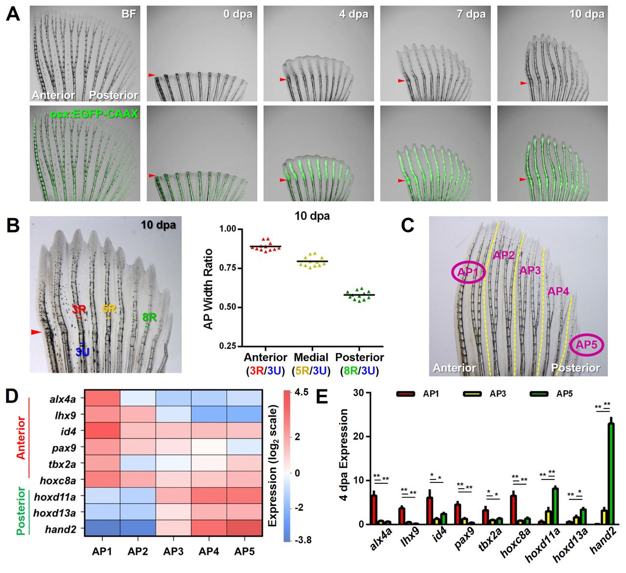
Fig. 1
Regeneration of pattern and underlying differential gene expression in pectoral fins. (A) Zebrafish pectoral fins possess and regenerate an anteroposterior (AP) skeletal pattern. Early osteoblasts marked by osx:EGFP-CAAX (green) are present in the regenerate prior to the onset of AP differences, which arise between 5 and 7 days post-amputation (dpa). Arrowheads indicate amputation plane. BF, bright field. (B) Zebrafish robustly regenerate the AP bone pattern. Quantification of the relative ratios between the width of the uninjured portion of anterior ray 3 (blue) and the widths of regenerating rays across the AP axis. n=12; bar indicates mean. (C) Diagram of a pectoral fin defining the two regions used for RNA-Seq (circled AP1 and AP5) and the five regions (AP1-AP5) used for subsequent qPCR validations. (D) Each AP region of uninjured zebrafish pectoral fins expresses a unique AP code of patterning transcription factor genes. Results shown reflect qPCR confirmation of RNA-Seq data, normalized to β-actin 1 (actb1) levels. n=3. (E) The AP regionalized expression of transcription factor genes is maintained during regeneration. qPCR expression profiles at 4 dpa, normalized to actb1 levels. n=3; mean ± s.e.m.; *P<0.05, **P<0.005, Student’s t-test.