
Image
|
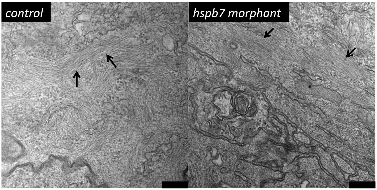
Figure Caption
Fig. S6
Myofibrils develop normally in embryos depleted of Hspb7. Shown are representative TEM images of frontal sections through the ventricles of 72 hpf embryos injected with 0.25 mM standard control MO (left, n = 2 embryos; 29 fields of view) or 0.25 mM hspb7 MO (right, n = 2 embryos, 31 fields of view). Bars indicate 200 nm. Arrows mark myofibrils.
Acknowledgments
This image is the copyrighted work of the attributed author or publisher, and
ZFIN has permission only to display this image to its users.
Additional permissions should be obtained from the applicable author or publisher of the image.
Reprinted from Developmental Biology, 381(2), Rosenfeld, G.E., Mercer, E.J., Mason, C.E., and Evans, T., Small heat shock proteins Hspb7 and Hspb12 regulate early steps of cardiac morphogenesis, 389-400, Copyright (2013) with permission from Elsevier. Full text @ Dev. Biol.