
|
Fig. 4
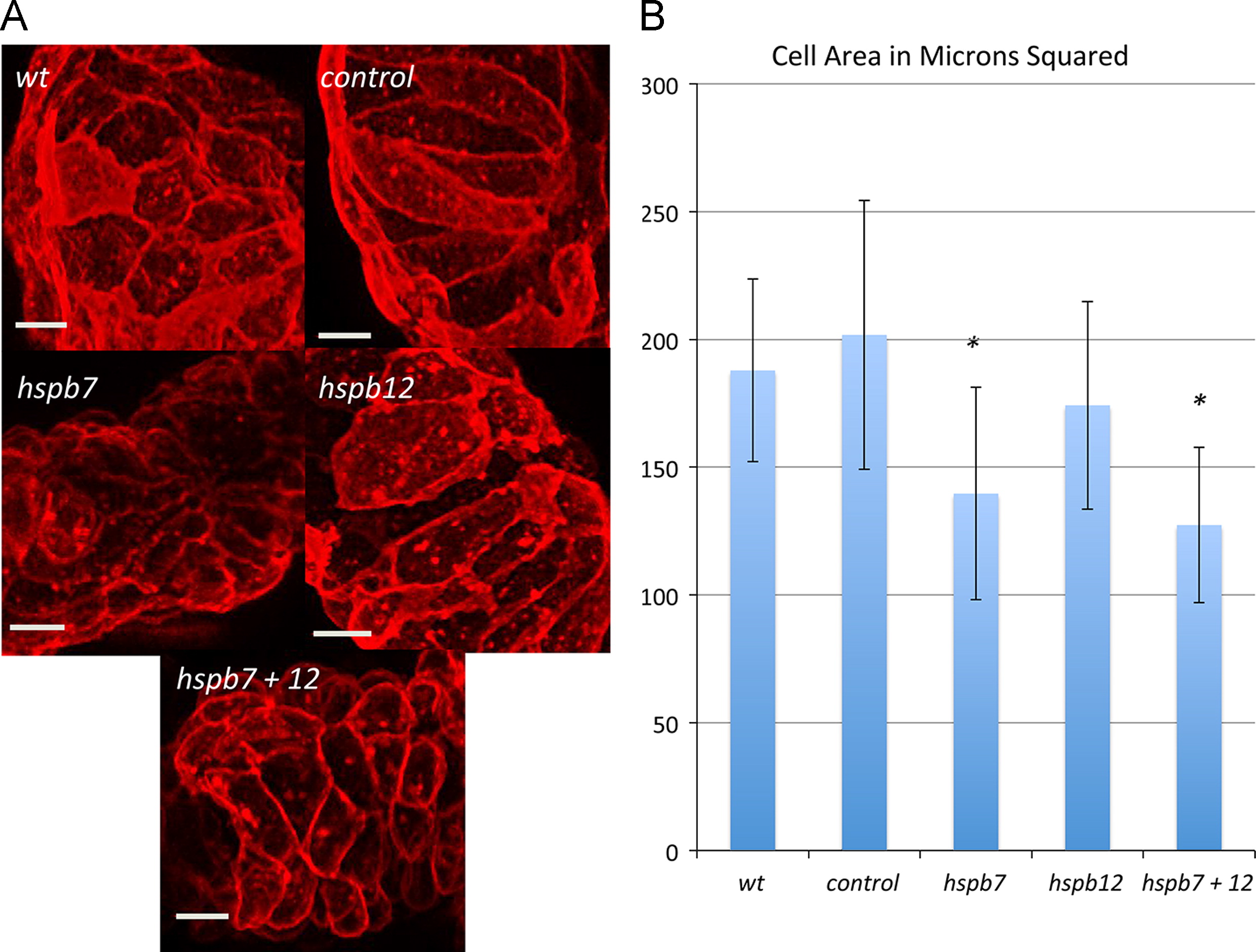
Hspb7 regulates ventricular cardiomyocyte size. (A) Representative phenotypes demonstrating the ventricular cell size observed in the indicated experimental conditions at 48 hpf in embryos derived from the myl7:actn3b; myl7:mKate-Caax reporter strain. The bars represent 10 μm. (B) Wildtype embryos (wt, n=3) or embryos injected with the standard control MO (control, n=4), hspb7 MO (hspb7, n=5), hspb12 MO (hspb12, n=3), or hspb7+12 MOs (hspb7+12, n=4) were evaluated with ImageJ to measure average cell area. At least seven cells were analyzed for each embryo to determine the average. The asterisk indicates p<0.05 by Student′s t-test in comparison to the standard control group.
Reprinted from Developmental Biology, 381(2), Rosenfeld, G.E., Mercer, E.J., Mason, C.E., and Evans, T., Small heat shock proteins Hspb7 and Hspb12 regulate early steps of cardiac morphogenesis, 389-400, Copyright (2013) with permission from Elsevier. Full text @ Dev. Biol.