
|
Fig. 2
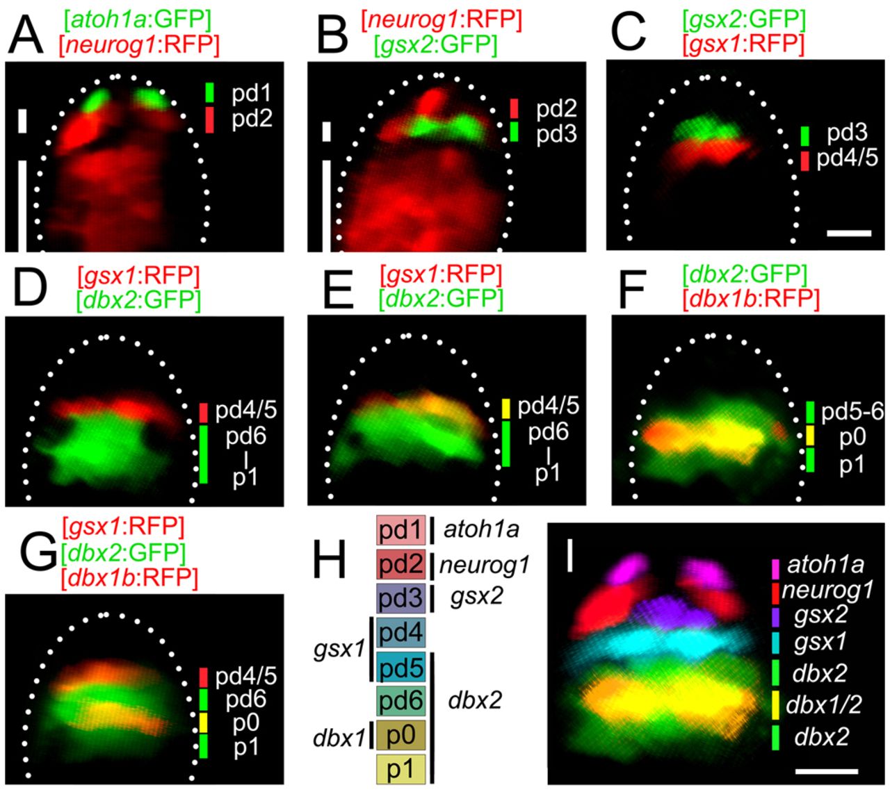
Progenitor domains in the dorsal spinal cord. Images were taken from embryos that were 36-48 hpf. All images are cross-sections of the spinal cord. Genotypes of the transgenic fish are shown at the top of each panel. (A) The atoh1a expression domain is located in the dorsal-most spinal cord. The short and long white lines show the dorsal and ventral neurog1 expression domains, respectively. The dorsal neurog1 expression domain is located just ventral to the atoh1a expression domain. (B) The gsx2 expression domain is located just ventral to the dorsal neurog1 expression domain. (C) The gsx2 and gsx1 expression domains are adjacent to each other. (D) The gsx1 and dbx2 expression domains are adjacent to each other. (E) In this image, the gsx1 expression domain is within the dbx2 expression domain (yellow). (F) The dbx1b expression domain is located in the middle of the dbx2 expression domain (yellow). (G) The gsx1 expression domain (red) and the dbx1b expression domain (yellow) are separate. (H) Summary of the domain organization. (I) Composite image of the expression domain of each transcription factor. For neurog1, only the dorsal expression domain is included. Scale bars: 10 μm.