
|
Fig. S6
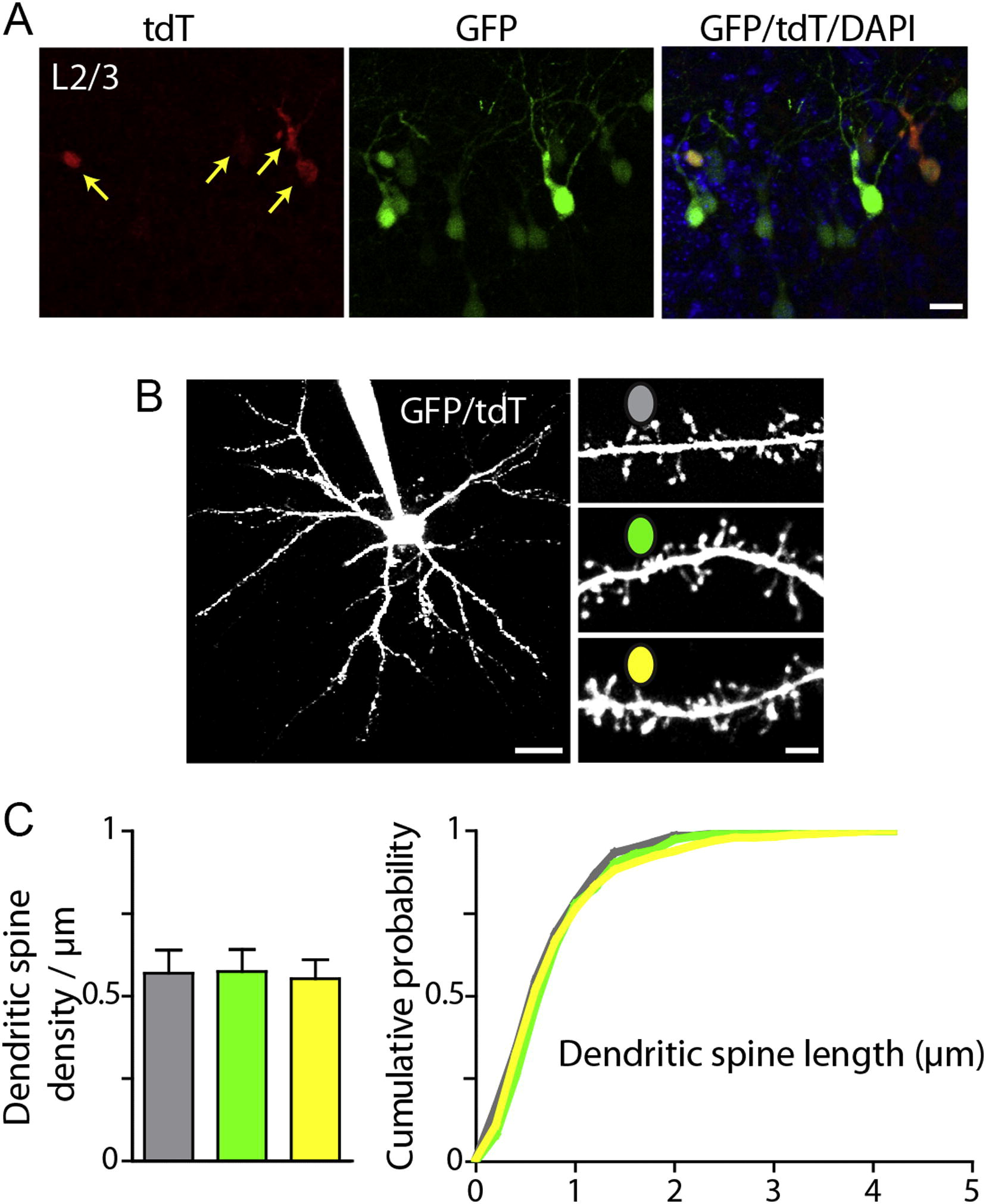
Structural Analysis of Electroporated Cortical Pyramidal Neurons, Related to Figure 5
Primary somatosensory cortex was electroporated at E15.5 with plasmids encoding GFP, ADG, DBDG, and UAS-tdT. T-DDOG is Gal4-GBP1p65-GBP6.
(A) Confocal image of native GFP and tdT fluorescence in layers 2/3 of mouse primary somatosensory cortex. Red, tdT; green, GFP; overlay shows both channels with DAPI. A subset of GFP+ cells are tdT+, reflecting co-electroporation of 4 constructs. Arrows point to GFP+/tdT+ cells. Scale bar, 20 μm.
(B) Left, 2-photon Z-stack through a GFP+/tdT+ layer 2/3 pyramidal neuron filled with Alexa-594 through a patch pipette. Right, dendrites from GFP-, GFP+/tdT- and GFP+/tdT+ neurons at P13. Scale bars, 30 and 3 μm.
(C) Summary data of dendritic spine density (left) and length (right) for electroporated pyramidal neurons at P12-14. Dendritic spine density and length did not differ among GFP-, GFP+/tdT- and GFP+/tdT+ neurons. (Dendritic spine density, n = 7 neurons and 15-24 dendrites per condition; spine length, <300 dendritic spines per condition). No GFP-/tdT+ neurons were observed in acute slices during recording or 2-photon imaging (n = 7 mice). Column plots are mean ± SEM.
Reprinted from Cell, 154(4), Tang, J.C., Szikra, T., Kozorovitskiy, Y., Teixiera, M., Sabatini, B.L., Roska, B., and Cepko, C.L., A nanobody-based system using fluorescent proteins as scaffolds for cell-specific gene manipulation, 928-939, Copyright (2013) with permission from Elsevier. Full text @ Cell