
Image
|
Figure Caption
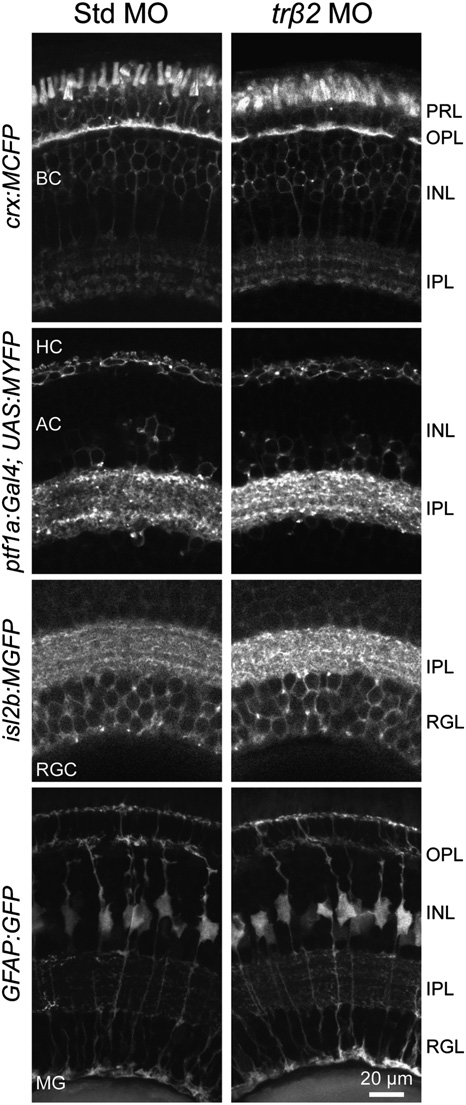
Fig. S7
Morphologies and distributions of the major retinal cell types in the trβ2 MO. Knocking down trβ2 does not affect the generation of bipolar cells (BC), HCs (HC), amacrine cells (AC), retinal ganglion cells (RGC), or Muller glia (MG), visualized using transgenic animals in which these major cell types are labeled by FP expression (12–14). Retinas from GeneTools standard control morphant (Std MO) and trβ2 morphant are shown. INL, inner nuclear layer; IPL, inner plexiform layer; OPL, outer plexiform layer; PRL, photoreceptor layer; RGL, retinal ganglion cell layer.
Acknowledgments
This image is the copyrighted work of the attributed author or publisher, and
ZFIN has permission only to display this image to its users.
Additional permissions should be obtained from the applicable author or publisher of the image.
Full text @ Proc. Natl. Acad. Sci. USA