
Image
|
Figure Caption
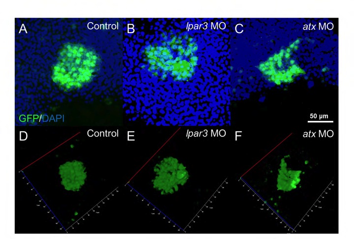
Fig. S5 KVs were deformed and dispersed in lpar3 and atx morphants. (A-F) GFP and DAPI merged images (A-C) and GFP image 3D stack model (D-F) of KV in control embryo (A), and lpar3 (B) and atx (C) morphants at the tail bud stage were examined and analyzed for cell dispersion and cell number under confocal microscopy. KV shown by GFP was from immunostaining of sox17:GFP embryos.
Acknowledgments
This image is the copyrighted work of the attributed author or publisher, and
ZFIN has permission only to display this image to its users.
Additional permissions should be obtained from the applicable author or publisher of the image.
Full text @ Development