
|
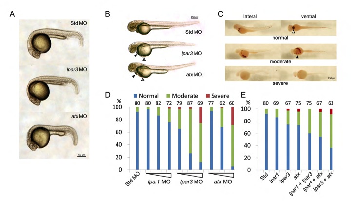
Fig. S3 Knockdown of atx and/or lpar3 causes cardiac edema and blood pooling. (A,B) Embryos injected with atx or lpar3 MOs were examined and photographed at 24 (A) and 48 (B) hpf. Cardiac edema (filled arrowheads) and blood pooling (open arrowheads) appeared at the ventral side of the yolk in both atx and lpar3 morphants were observed (B). (C) Embryos injected with atx or lpar3 MOs were fixed and stained with o-dianosidine to reveal erythrocytes (brown staining). The treated embryos were positioned and photographed from the lateral view or ventral view. In the normal condition (top row), erythrocytes concentrated in pericardiac cavity (open arrowhead) and major vessels; however, in moderate defective embryos (middle row), erythrocytes spread out to the ventral side of the yolk (filled arrowhead); in the most severe condition (lower row), were reduced erythrocytes and hemorrhage in brain and/or eyes were sometimes observed (ventral view). (D) Dose-dependent disturbance of erythrocyte distribution was found in embryos injected with 1.25, 2.5 and 5 ng per embryo of lpar1, lpar3 or atx MOs. Std MO at 5 ng was used as a control (n=3). (E) Embryos were injected with single or different combinations of 1.25 ng of the indicated MOs.