
Image
|
Figure Caption
Fig. 1
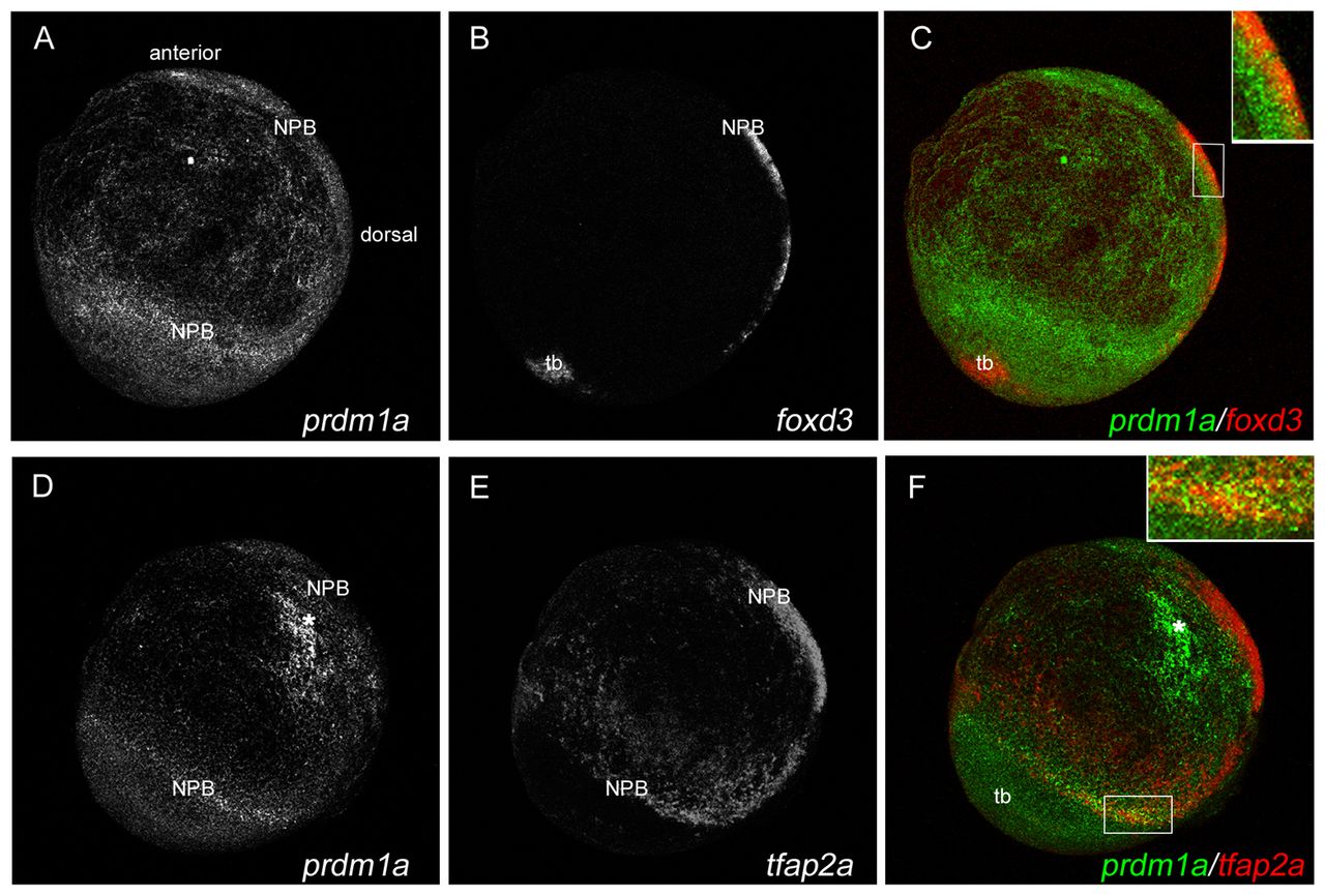
prdm1a is co-expressed with foxd3 and tfap2a at the NPB. Confocal micrograph projections of double fluorescent in situ hybridization (ISH) of 2-somite (11 hpf) wild-type (WT) zebrafish embryos for prdm1a with foxd3 (A-C) and for prdm1a with tfap2a (D-F). prdm1a (green) is co-expressed with both foxd3 (red, C) and tfap2a (red, F) at the NPB, as represented in yellow in the merged images (see insets in C and F). All images are lateral views with anterior to the top, dorsal to the right. Asterisk indicates non-specific staining. NPB, neural plate border; tb, tailbud.
Acknowledgments
This image is the copyrighted work of the attributed author or publisher, and
ZFIN has permission only to display this image to its users.
Additional permissions should be obtained from the applicable author or publisher of the image.
Full text @ Development