
Image
|
Figure Caption
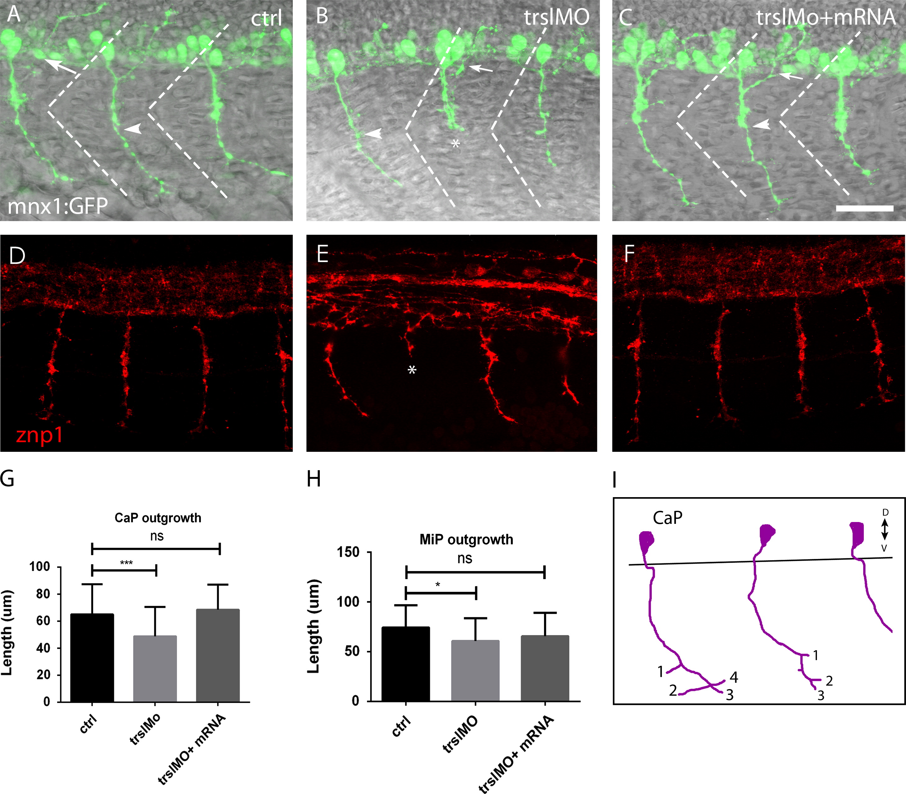
Fig. 5 Defective primary motor neurons. PMNs in the spinal cord of control embryos (A), trslMO (B) and trslMO +mRNA (C) injected Tg(mnx1:GFP) embryos (B) have dorsally projecting MiPs (arrow) and ventrally projecting CaP (arrowhead). Staining of control (D), trslMO (E) and trslMO +mRNA (F) injected embryos using znp1. CaP neurons stalling was observed in trslMO injected embryos (N). Measurement of the length of CaP (G) and MiP (H). Bar is 20 μm. Two-way ANOVA test: NNNP<0.0005, NP<0.05, n.s.=not significant.
Figure Data
Acknowledgments
This image is the copyrighted work of the attributed author or publisher, and
ZFIN has permission only to display this image to its users.
Additional permissions should be obtained from the applicable author or publisher of the image.
Reprinted from Developmental Biology, 381(2), Abramsson, A., Kettunen, P., Banote, R.K., Lott, E., Li, M., Arner, A., and Zetterberg, H., The zebrafish amyloid precursor protein-b is required for motor neuron guidance and synapse formation, 377-88, Copyright (2013) with permission from Elsevier. Full text @ Dev. Biol.