
Image
|
Figure Caption
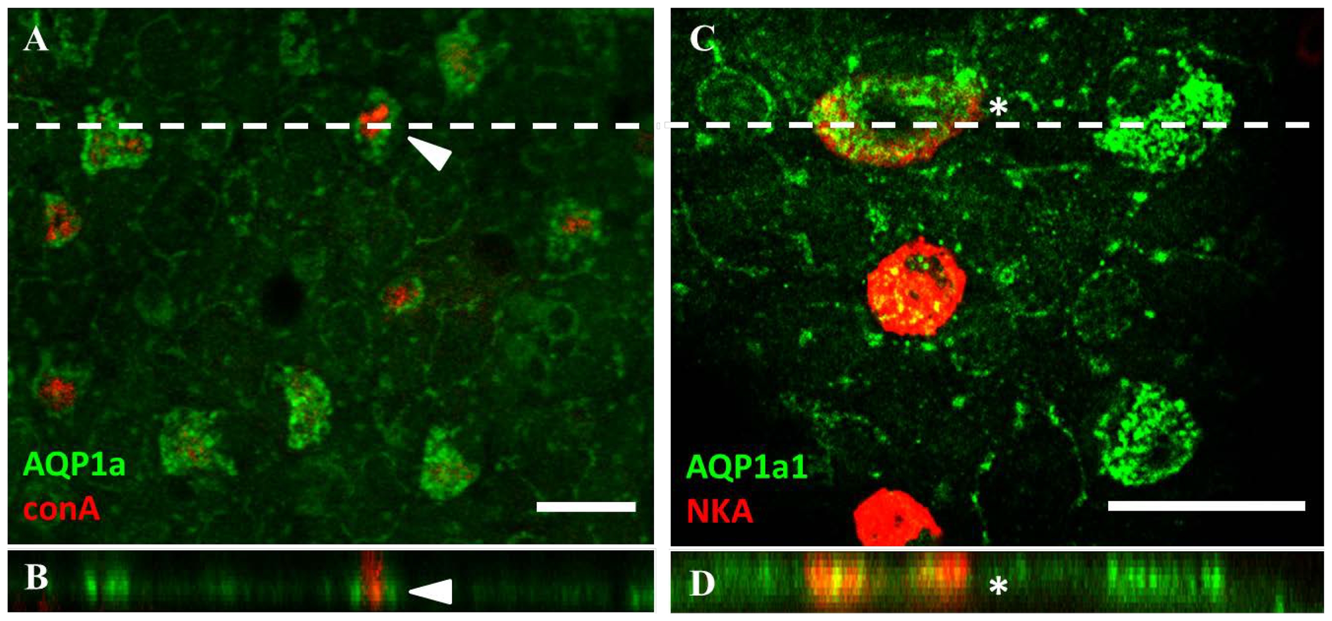
Fig. 4
Aquaporin-1a1 is expressed predominantly on the basolateral membrane of ionocytes.
Fluorescent immunohistochemistry and z-stack analysis with confocal microscopy revealed that (A and B) the expression of aquaporin-1a1 (AQP1a1; green) does not overlap with concanavalin-A staining (arrow; red). (C and D) However, AQP1a1 expression is found to co-localize with Na+/K+-ATPase (NKA; red) staining (cell labelled with an asterisk). Dashed lines in A and C indicate the position for z-stack images in B and D, respectively. Scale bar = 20 μm.
Figure Data
Acknowledgments
This image is the copyrighted work of the attributed author or publisher, and
ZFIN has permission only to display this image to its users.
Additional permissions should be obtained from the applicable author or publisher of the image.
Full text @ PLoS One