
|
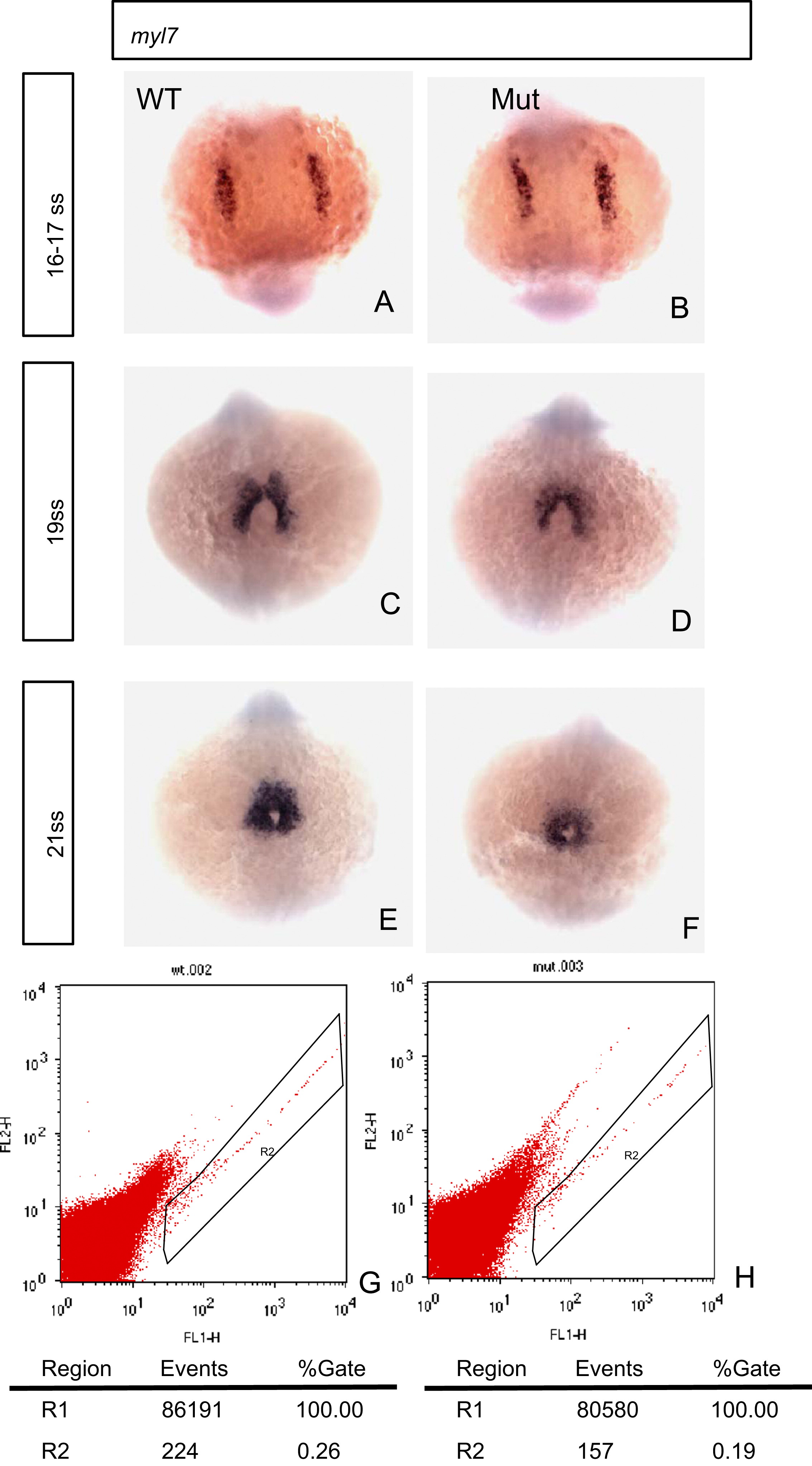
Fig. 3 Heart development is affected in the eif3ba mutant as shown by myl7 in situ hybridization and FACS analyses. (A, B) At 16–17 ss, the development of the cardiomyocytes appears normal in the mutant compared with the wild-type. (C, D) At 19 ss, the boundary of the myl7 expression region is wider and the signal is stronger in the wild-type than in the mutant. (E, F) At 20 ss, the myocardium is thicker in the wild-type than in the mutant. (G, H) FACS analysis of the EGFP-positive cells of both wild-type and mutant embryos (20 embryos in each group, 28 hpf), on the background of the Tg(myl7:EGFP) fish line. The number of EGFP-positive cardiomyocytes in the mutant is significantly fewer than that in the wild-type. WT, wild type; Mut, eif3ba mutant. (A)–(F) The embryos are in dorsal view with anterior down.
Reprinted from Developmental Biology, 381(1), Xia, Z., Tong, X., Liang, F., Zhang, Y., Kuok, C., Zhang, Y., Liu, X., Zhu, Z., Lin, S., and Zhang, B., Eif3ba regulates cranial neural crest development by modulating p53 in zebrafish, 83-96, Copyright (2013) with permission from Elsevier. Full text @ Dev. Biol.