
|
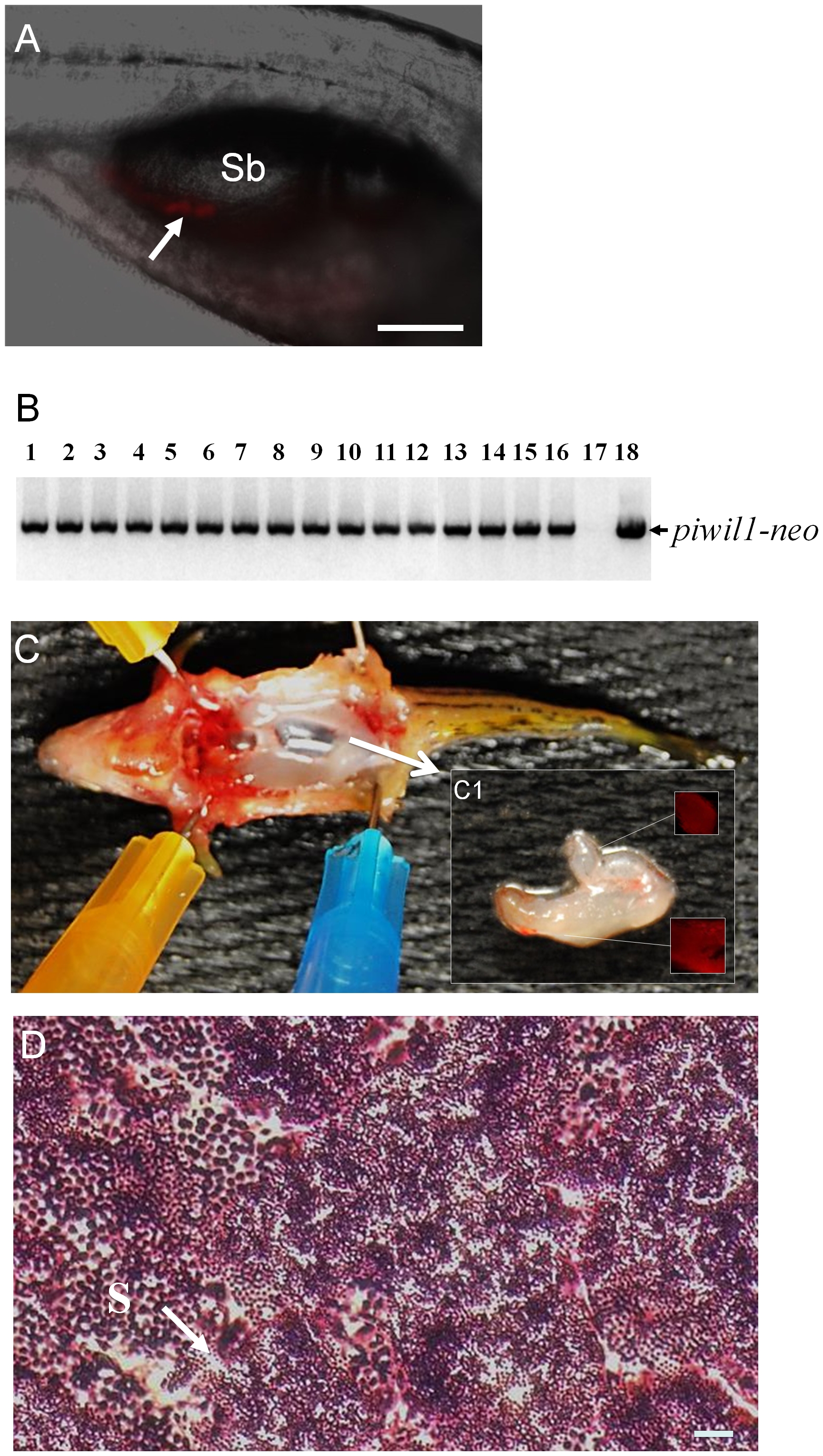
Fig. 6
Germline transmission of spermatogonia cultured for 6 weeks in the presence of dorsomorphin.
(A) Photomicrograph showing the incorporation of DsRed-positive cultured spermatogonia (arrow) into the gonad of a recipient larva two weeks after transplantation. (B) Results of genomic PCR showing the presence of piwil1-neo sequences that were inherited by all of the F1 individuals (lanes 1 to 16) produced by a germline chimeric father. Negative control: genomic DNA template from a wild-type larva (lane 17); positive control: pPiwil1-neo plasmid DNA template (lane 18). (C) Dissection of a fertile adult male recipient fish showing that the transplanted DsRed-positive spermatogonia have proliferated and directed the formation of a pair of unequal sized testes (arrow) in the body. (C1) Inset shows the gonad under UV light revealing the presence of DsRed-positive cells. (D) Transverse section of testis from a fertile recipient fish showing active spermatogenesis. Sb: swim bladder; S: spermatozoa (arrow). Scale bar = 100 μm for A and 20 μm for D.