
|
Fig. 5
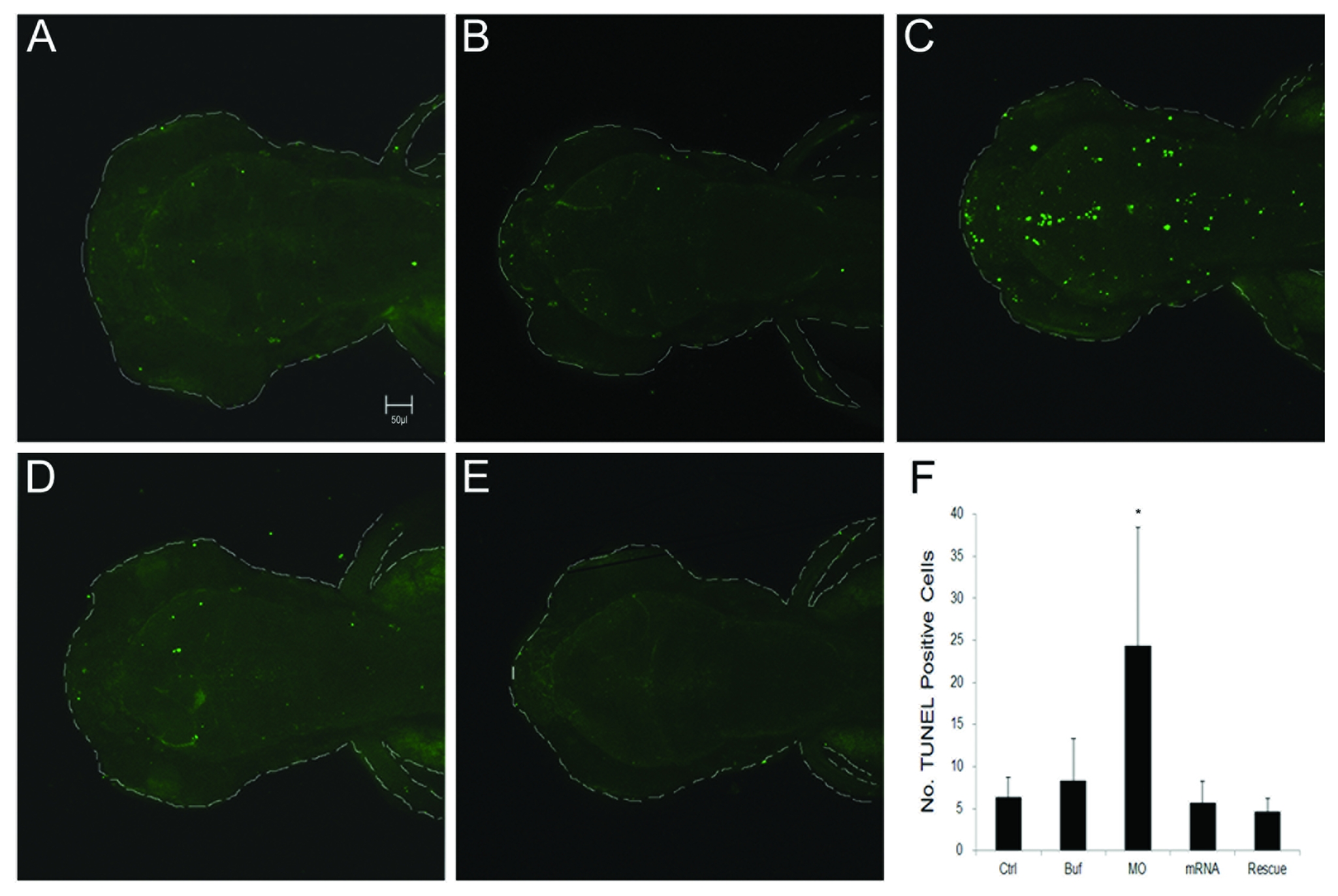
TUNEL staining for apoptosis in treated larvae.
All larvae were evaluated at 3 dpf. (A) Control. (B) Injection of 1x Danieaus’ buffer with 1% phenol red. (C) Injection of 5 ng ddc tMO1. (D) Injection of 100 pg ddc mRNA. (E) Injection of mixture of 5 ng ddc tMO1 and 50 pg ddc mRNA with non-MO1 binding site. Apoptotic cells were increased in ddc morphants compared to other controls. The increase of apoptosis can be attenuated by co-injection of ddc mRNA. (F) Statistical analysis. Photographs were taken by Zeiss Confocal 700 microscope at 100X magnification. All data represented at least three independent experiments and were expressed as mean±SD. *p<0.05 compared to control larvae.