
Fig. 9
- ID
- ZDB-IMAGE-131009-27
- Genes
- Publication
- Drummond et al., 2013 - The role of Zic transcription factors in regulating hindbrain retinoic acid signaling
- All Figures
- Figures for Drummond et al., 2013
|
Fig. 9
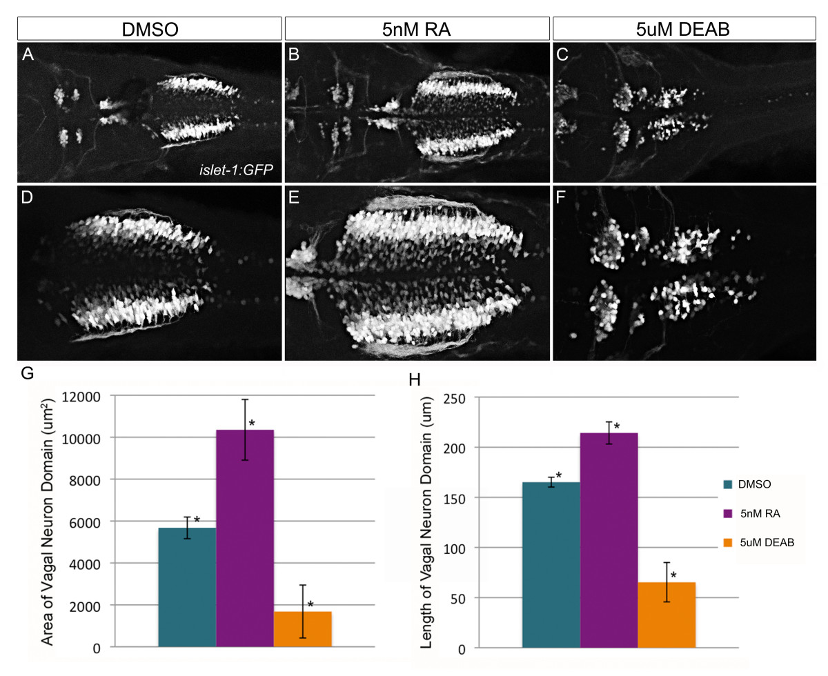
Vagal neurons are sensitive to alterations in retinoic acid levels.Tg(isl1:eGFP) transgenic zebrafish are used to visualize alterations to branchiomotor neurons in response to 1% DMSO (A, D), 5 nM all-trans retinoic acid (B, E), or 5 μM DEAB (C, F) treatment. Images are dorsal views of 48 hpf embryo hindbrain using 20× or (A-C) or 40× (D-F) objective. Length (G) and area (H) were quantified using ImageJ and displayed graphically (error bar denotes standard deviation). The differences induced by either retinoic acid or DEAB treatment are statistically significant (ANOVA and Tukey’s HSD post-hoc test, *p-value < 0.01).