Image


                        Figure Caption


Fig. 3
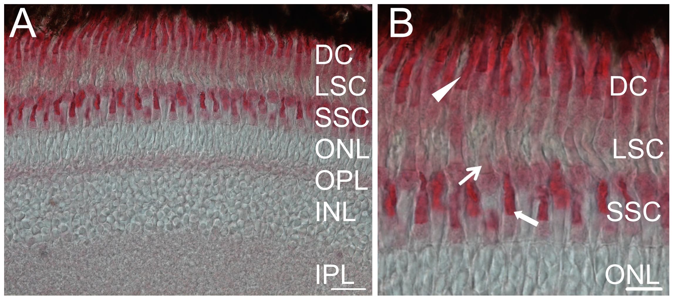
Immunostaining of zGC3 in cryosections of adult zebrafish retina.
Sectionas were probed with the anti-zGC3 antibody (1:400). The secondary antibody was used at 1:200 dilution. (A) Section of a whole retina, cell layers are indicated as double cones (DC), long single cones (LSC), short single cones (SSC), outer nuclear layer (ONL), outer plexiform layer (OPL), inner nuclear layer (INL) and inner plexiform layer (IPL). (B) Magnified part of (A). DC: closed arrowhead; LSC: thin arrow; SSC: bold arrow. Scale bar = 20 μm in (A) and 10 μM in (B).
                
                    
                        Figure Data


                        Acknowledgments


This image is the copyrighted work of the attributed author or publisher, and
ZFIN has permission only to display this image to its users.
Additional permissions should be obtained from the applicable author or publisher of the image.
      Full text @ PLoS One


        