
Image
|
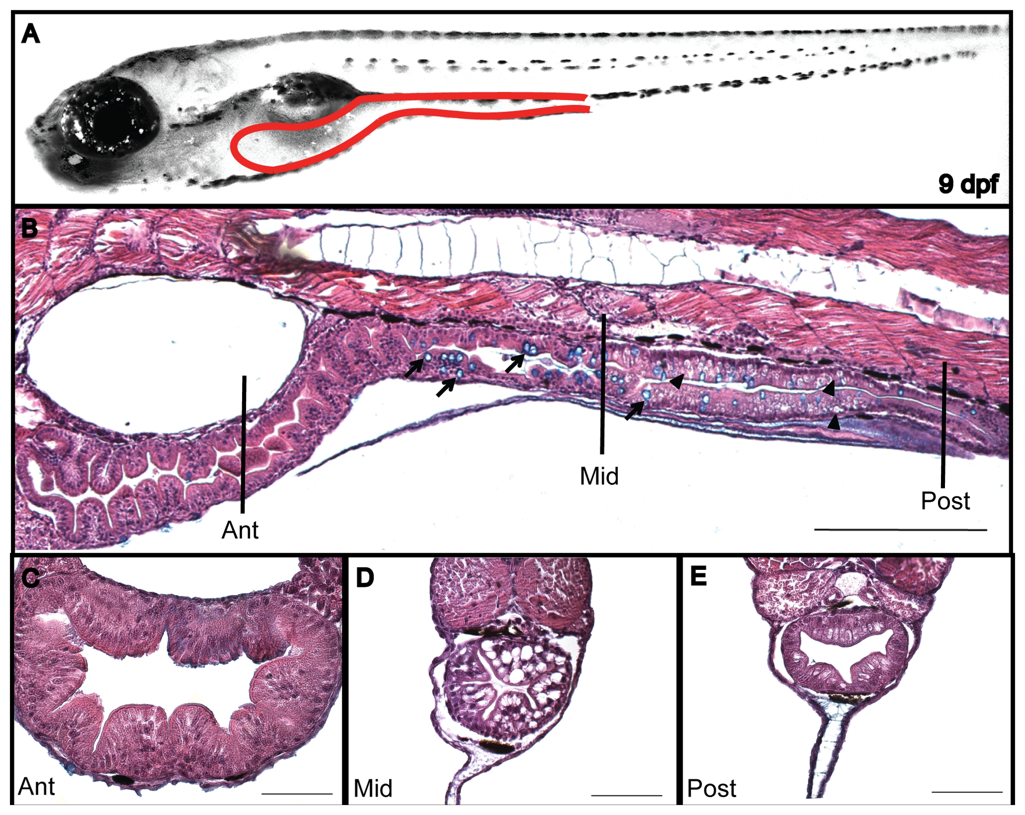
Figure Caption
Fig. 4
Intestine morphology of a 9dpf larva.
(A) Larva lateral view with the intestine delineated (dotted red line). Sagittal (B) and transversal (C–E) paraffin sections stained with H&E and alcian blue. The intestine is divided in 3 different segments: anterior (C) middle (D) and posterior (E). The arrow indicates goblet cells and the arrowhead indicates a supranuclear vesicle. Scale bar = 200μm in B; 50µm in C, D and E.
Acknowledgments
This image is the copyrighted work of the attributed author or publisher, and
ZFIN has permission only to display this image to its users.
Additional permissions should be obtained from the applicable author or publisher of the image.
Full text @ PLoS One