
|
Fig. 3
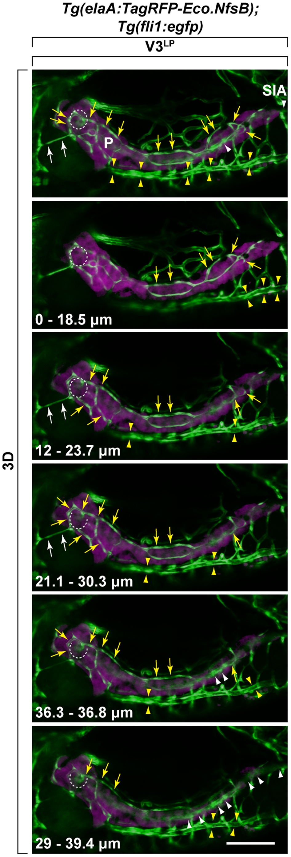
Identification of V3LP vessel that bridges between liver and pancreas.
V3LP vessel originates from supraintestinal arteries (SIA). The origins of V3LP was followed by using Tg(elaA:TagRFP-Eco.NfsB);Tg(fli1:egfp) at 5 dpf. The 3D-rendered Z-stack confocal miscroscopy images of several Z-slices (depth of ranges is indicated at the bottom left in each panel) are shown in series. Fli1+ vessels and elaA+ exocrine pancreas are shown as green and magenta, respectively. By following SIA (white arrowheads) in each Z-stack, V3LP is found to be connected to a dorsal SIA branch (yellow arrows), to vascular plexus at the islet of Langerhans (dotted circle) that is linked to both dorsal (yellow arrows) and ventral (white arrowheads) SIA branches, and to SIV (sandwiched between yellow arrowheads), a part of which appears to be embedded inside pancreas. Supraintestinal artery (SIA): White arrowheads; Supraintestinal vein (SIV): Sandwiched between yellow arrowheads. Scale bars: 100 μm.