
|
Fig. 1
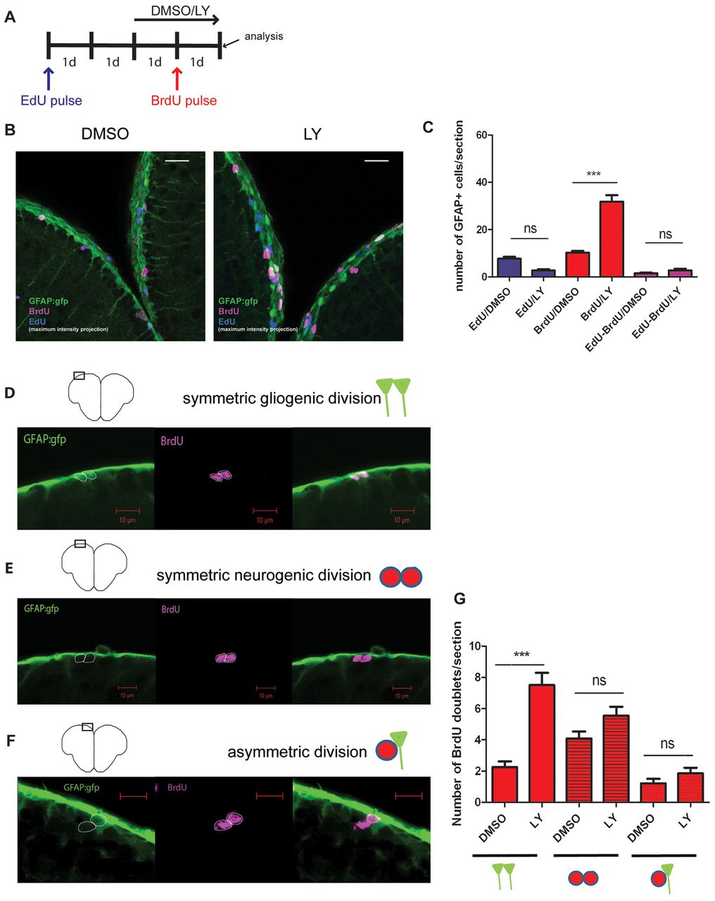
Symmetrically dividing radial glial cells (RG) are recruited into the cell cycle during Notch blockade. (A) Experimental design. An initial cohort of RG in S phase was labeled with an EdU pulse followed by a second pulse of BrdU during Notch blockade (LY). 1d, 1-day intervals. (B) Triple immunostaining showing GFAP- (green), BrdU- (magenta) and EdU- (blue) positive cells in the Dm region of the pallium (confocal projections over 16 and 19 μm, respectively). (C) Quantification of EdU-positive, BrdU-positive and EdU/BrdU-positive RG. Red bars, P<0.0001; blue bars, P=0.07; pink bars, P>1 (n=4 brains for each treatment, total number of cells counted: 1156). (D-G) Division modes of RG and quantification of BrdU-positive doublets in the experimental setting shown in A. Symmetric gliogenic divisions, P<0.0001; other division modes, P>1; n=273 doublets counted. Scale bars: white, 20 μm; red 10 μm.