
|
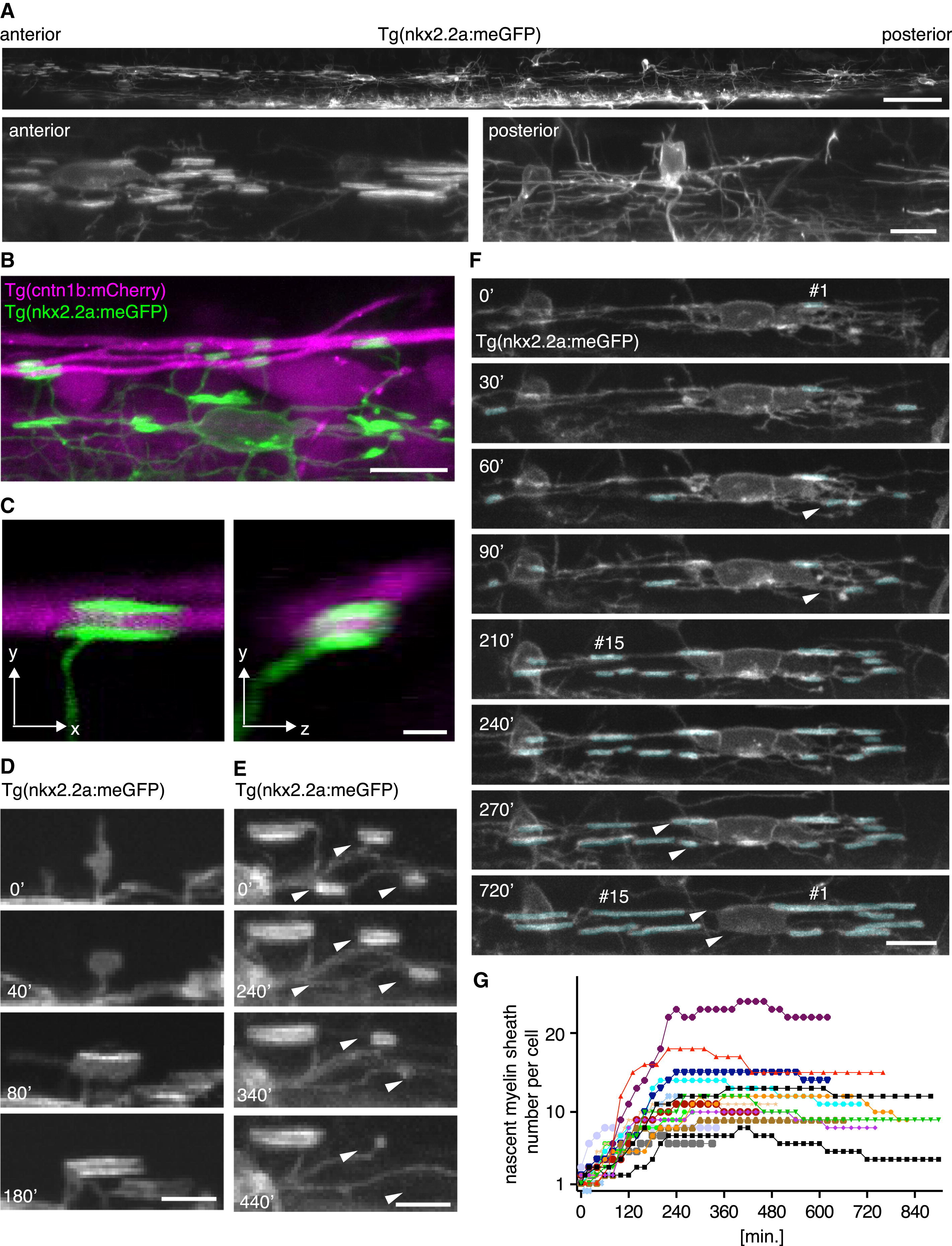
Fig. 2
Individual Oligodendrocytes Initiate New Myelin Sheaths in a Short Time Window (A) Top: lateral view of a 0.75 mm stretch of the zebrafish spinal cord at 3 dpf indicates a gradient from anterior to posterior in the differentiation status of oligodendrocytes in a Tg(nkx2.2a:meGFP) animal. Scale bar, 50 μm. Bottom: cells located in the anterior spinal cord have a mature myelinating morphology (left), whereas those in more posterior regions of the spinal cord have a less mature premyelinating morphology (right). Scale bar, 10 μm. (B) Lateral view of a Tg(nkx2.2a:mEGFP), Tg(cntn1b:mCherry) double transgenic zebrafish at 3 dpf shows a single oligodendrocyte with nascent myelin sheaths surrounding axons. Scale bar, 10 μm. (C) Higher magnification x,y (left panel) and x,z (right panel) views of a Tg(nkx2.2a:mEGFP), Tg(cntn1b:mCherry) animal confirm that nkx2.2a:mEGFP-labeled oligodendrocyte processes surround axons. Scale bar, 2 μm. (D) Single selected images from a time-lapse series of an oligodendrocyte process in a Tg(nkx2.2a:mEGFP) zebrafish reveal transformation of an exploratory process into a myelin sheath within a few hours. Scale bar, 5 μm. (E) Single selected images from a time-lapse series showing that nascent myelin sheaths can be retracted (arrowheads) over a period of a few hours. Scale bar, 5 μm. (F) Selected images from a time-lapse series of a single oligodendrocyte in a Tg(nkx2.2a:mEGFP) animal. The cell makes its first nascent myelin sheath (indicated by “#1”) at time point zero (see main text) and its final new myelin sheath (indicated by “#15”) by the 210 min time point. Examples of nascent myelin sheaths that are retracted during the time-lapse are indicated by arrowheads. Scale bar, 10 μm. (G) Total number of nascent myelin sheaths for 16 different oligodendrocytes over time. Time point zero is the time at which each oligodendrocyte initiates its first myelin sheath. The cell from (F) is colored in cyan. See also Movie S1.
Reprinted from Developmental Cell, 25(6), Czopka, T., Ffrench-Constant, C., and Lyons, D.A., Individual Oligodendrocytes Have Only a Few Hours in which to Generate New Myelin Sheaths In Vivo, 599-609, Copyright (2013) with permission from Elsevier. Full text @ Dev. Cell