
|
Fig. 5
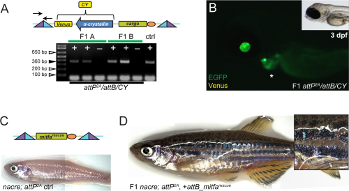
Tissue-specific promoter transgenes created using phiC31 integrase. A,B: pDestattB/CY, containing α-crystallin:Venus (CY, yellow fluorescence) as transgenesis marker (schematic), conveys eye lens-specific transgene expression when stably integrated into attP landing sites; the agarose gel illustrates positive (+) F1 transmission as detected by PCR from 2 representative independent F1 founders (A and B) including negative control siblings (), primers for attL indicated in schematic. Asterisk in B indicates EGFP expression from cmlc2:EGFP. C,D: Transgenics with an attB_mitfarescue transgene in mitfa-mutant nacre containing the attP2A site; (C) schematic of the final transgene integration and initial adult phenotype without melanocytes in uninjected control (ctrl), compared to (D) adult transgenic with stable F1 attP2A/attB_mitfarescue integration (inset shows skin close-up).