
|
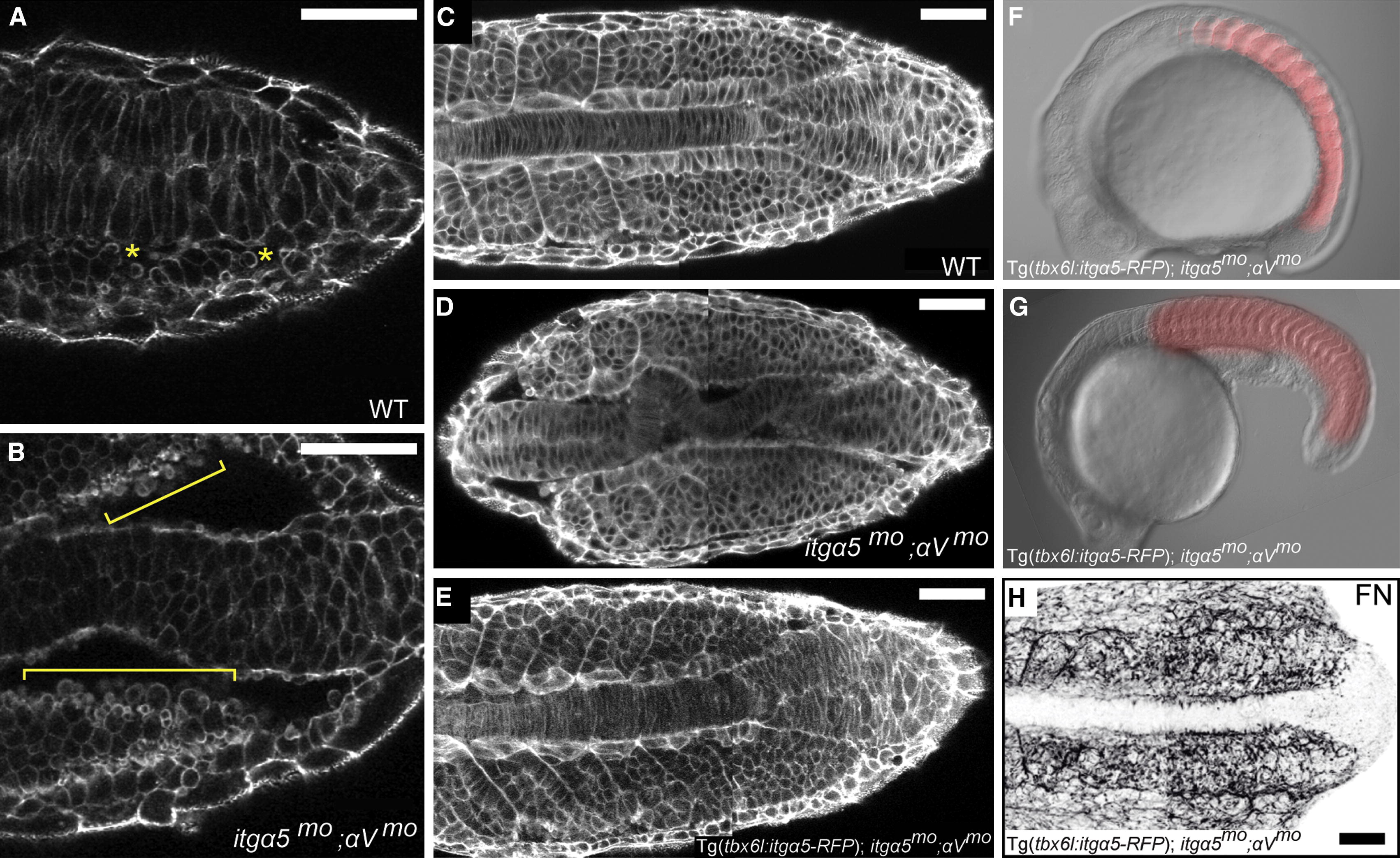
Fig. 4
itgα5 Function in the Paraxial Mesoderm Is Sufficient to Rescue Body Elongation in itgα5mo;αVmo Embryos (A) In WT embryos, cells on the surface of the paraxial mesoderm display low levels of blebbing (asterisks), as revealed by phalloidin staining. (B) In itgα5mo;αVmo embryos, cells along the medial surface of the paraxial mesoderm exhibit a dramatic increase in blebbing (brackets). See also Movie S2. (C–E) Phalloidin staining of the cell cortices shows the close alignment of the notochord and paraxial mesoderm in WT embryos (C) (n = 16), the loss of this intertissue adhesion and alignment in itgα5mo;αVmo embryos (D) (n = 31), and the rescue of intertissue adhesion and organization in Tg(tbx6l:itgα5-RFP);itgα5mo;αVmo embryos (E) (n = 10). (F and G) Body elongation is rescued in Tg(tbx6l:itgα5-RFP);itgα5mo;αVmo embryos at 14 hpf (F) and 24 hpf (G). At the 16-somite stage, the trunks of transgenic rescue embryos (n = 40) are 88% of that in WT embryos (SD = 4%; p < 0.05), and the heads are 83% of that in WT embryos (SD = 5%; p < 0.05). RFP fluorescence shows Itgα5 expression in the paraxial mesoderm. (H) Tg(tbx6l:itgα5-RFP) rescues FN matrix assembly and fiber orientation in itgα5mo;αVmo embryos (n = 7). Anterior is left. Note that panels (C–E) and (H) are composites of anterior and posterior images. Scale bars represent 50 µm. See also Figure S3.